 Back
BackThe Brain and Cranial Nerves: Structure, Function, and Organization
Study Guide - Smart Notes
 Tailored notes based on your materials, expanded with key definitions, examples, and context.
Introduction to the Brain and Cranial Nerves
The adult human brain is the central organ of the nervous system, containing nearly 97% of the body's nervous tissue. It is responsible for processing sensory information, coordinating movement, and higher mental functions such as thought, memory, and emotion. The brain is divided into several major regions, each with specialized functions.
Average weight: 1.4 kg (3 lb)
Volume: Typically 1200 mL (range: 750–2100 mL)
Note: Brain size does not correlate with intelligence.
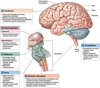
The Brain: Major Regions and Their Functions
Cerebrum
The cerebrum is the largest part of the adult brain, responsible for conscious thought, sensation, intellect, memory, and complex movements. It is divided into left and right hemispheres, each covered by a highly folded layer of gray matter called the cerebral cortex.
Gyri: Rounded elevations that increase surface area
Sulci: Shallow grooves between gyri
Fissures: Deeper grooves separating major regions
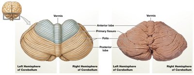
Cerebellum
The cerebellum coordinates ongoing body movements and helps maintain balance and posture. It consists of two hemispheres and is covered by the cerebellar cortex (gray matter).
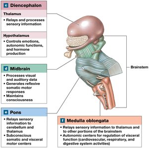
Diencephalon
Thalamus: Relays and processes sensory information
Hypothalamus: Controls emotions, autonomic functions, and hormone production; integrates nervous and endocrine systems
Brainstem
Midbrain: Processes visual and auditory information; maintains consciousness
Pons: Relays information to/from cerebellum; involved in motor control
Medulla oblongata: Relays sensory information; regulates autonomic functions (heart rate, blood pressure)
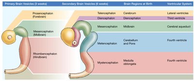
Embryonic Development of the Brain
The central nervous system develops from a neural tube, with the cephalic portion forming three primary brain vesicles that further subdivide into secondary vesicles and mature brain regions.
Primary Vesicle | Secondary Vesicle | Adult Brain Region | Ventricular System |
|---|---|---|---|
Prosencephalon (Forebrain) | Telencephalon | Cerebrum | Lateral ventricles |
Diencephalon | Diencephalon | Third ventricle | |
Mesencephalon (Midbrain) | Mesencephalon | Midbrain | Cerebral aqueduct |
Rhombencephalon (Hindbrain) | Metencephalon | Cerebellum and Pons | Fourth ventricle |
Myelencephalon | Medulla oblongata | Fourth ventricle |
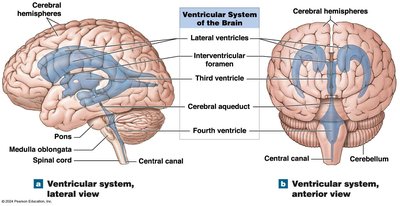
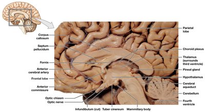
Ventricular System of the Brain
The brain contains four ventricles—fluid-filled chambers lined with ependymal cells—that produce and circulate cerebrospinal fluid (CSF).
Lateral ventricles: One in each cerebral hemisphere, separated by the septum pellucidum
Third ventricle: In the diencephalon, connected to lateral ventricles via interventricular foramen
Fourth ventricle: Extends into the medulla oblongata, connects to the central canal of the spinal cord and third ventricle via the cerebral aqueduct
Brain Protection and Support
Physical Protection
Bones of the skull
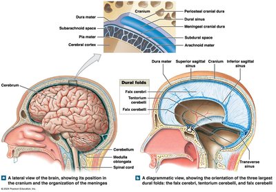
Cranial meninges: Three layers—dura mater (outer), arachnoid mater (middle), pia mater (inner)
Cerebrospinal fluid (CSF): Cushions and supports the brain
Cranial Meninges
Dura mater: Outermost, tough layer; forms dural folds (falx cerebri, tentorium cerebelli, falx cerebelli) that stabilize the brain
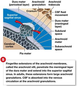
Arachnoid mater: Middle, web-like layer; subarachnoid space contains CSF
Pia mater: Innermost, delicate layer attached to brain surface
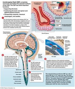
Cerebrospinal Fluid (CSF)
CSF surrounds all exposed surfaces of the CNS, supporting and protecting the brain, and transporting nutrients, chemical messengers, and wastes.
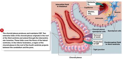
Formation: Produced by the choroid plexus in each ventricle by specialized ependymal cells
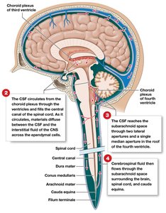
Circulation: Flows from the choroid plexus through ventricles, central canal, and subarachnoid space; absorbed into venous circulation via arachnoid granulations
Blood-Brain Barrier (BBB) and Blood Supply
The BBB isolates the CNS from general circulation, allowing selective transport of substances. It is formed by capillary endothelial cells with tight junctions, regulated by astrocytes. The blood-CSF barrier, formed by ependymal cells, limits transfer of substances to CSF. Breaks in the BBB occur in circumventricular organs for hormone release.
Blood supply: Internal carotid and vertebral arteries supply the brain; internal jugular veins remove blood
Cerebrovascular accident (stroke): Interruption of blood flow causes neuron death
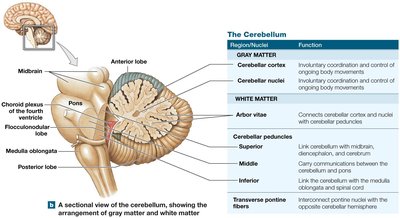
The Brainstem: Medulla Oblongata, Pons, and Midbrain
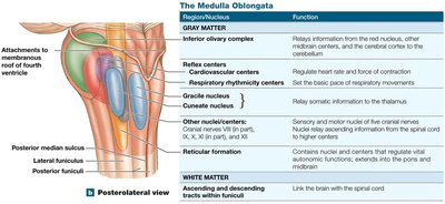
Medulla Oblongata
The medulla oblongata is the most inferior part of the brainstem, containing nuclei for autonomic reflexes, cranial nerves, and sensory/motor relay stations.
Reflex centers: Cardiovascular and respiratory rhythmicity centers
Relay stations: Gracile and cuneate nuclei (somatic sensory), solitary nuclei (visceral sensory), inferior olivary complex (motor coordination)
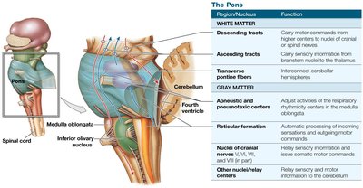
Pons
The pons contains nuclei for respiration control, relays information to/from the cerebellum, and contains ascending, descending, and transverse fibers.
Respiratory centers: Apneustic and pneumotaxic centers
Relay nuclei: Link cerebellum with other brain regions
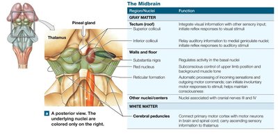
Midbrain
The midbrain processes visual and auditory information, maintains consciousness, and contains motor nuclei.
Tectum: Superior colliculi (visual reflexes), inferior colliculi (auditory reflexes)
Tegmentum: Red nucleus (motor commands), substantia nigra (dopamine production)
Cerebral peduncles: Motor fibers connecting cerebrum and brainstem
The Cerebellum
The cerebellum coordinates and fine-tunes voluntary and involuntary movements, maintains posture and balance, and stores learned motor patterns.
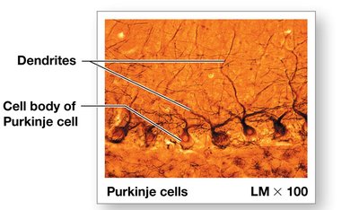
Cerebellar cortex: Gray matter with folia (folds) and Purkinje cells
Arbor vitae: Internal white matter connecting cortex and nuclei
Cerebellar peduncles: Superior, middle, and inferior tracts linking cerebellum to other brain regions
Disorders: Ataxia (loss of coordination), often due to trauma, stroke, or alcohol
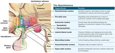
The Diencephalon
The diencephalon integrates sensory information and motor commands at a subconscious level. It includes the epithalamus, thalamus, and hypothalamus.
Epithalamus: Contains the pineal gland (secretes melatonin for circadian rhythms)
Thalamus: Relays and filters sensory information to the cerebral cortex; involved in motor and sensory integration
Hypothalamus: Regulates autonomic functions, emotions, hormone production, and links nervous and endocrine systems
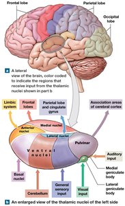
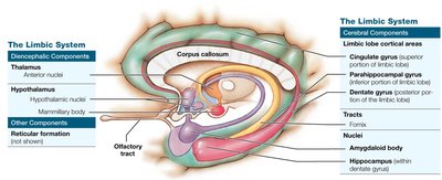
The Limbic System
The limbic system is a functional grouping of brain structures involved in emotion, motivation, and memory. It links conscious and unconscious brain functions and facilitates memory storage and retrieval.
Components: Limbic lobe (cingulate, dentate, parahippocampal gyri), hippocampus, amygdaloid body, fornix, anterior thalamic nuclei, hypothalamus
Functions: Establishes emotional states, links conscious and autonomic functions, facilitates memory
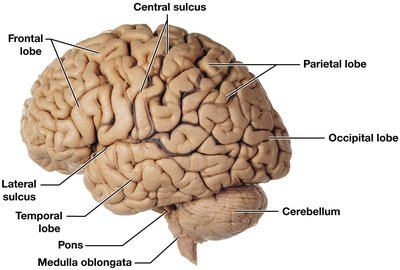
The Cerebrum: Structure and Function
The cerebrum is responsible for conscious thought, intellectual functions, and processing somatic sensory and motor information. It consists of gray matter (cerebral cortex, basal nuclei) and white matter (deep to cortex).
Gyri: Elevations increasing cortical surface area
Sulci: Grooves separating gyri
Lobes: Frontal, parietal, temporal, occipital, insula
White Matter of the Cerebrum
Association fibers: Connect areas within one hemisphere
Commissural fibers: Connect the two hemispheres (e.g., corpus callosum)
Projection fibers: Link cortex to lower brain regions and spinal cord
Basal Nuclei
The basal nuclei (basal ganglia) are masses of gray matter deep within the cerebral hemispheres. They modulate voluntary motor commands and muscle tone.
Caudate nucleus
Lentiform nucleus: Putamen (lateral), globus pallidus (medial)
Functional Areas of the Cerebral Cortex
Primary motor cortex: Controls voluntary skeletal muscles (precentral gyrus, frontal lobe)
Primary somatosensory cortex: Receives touch, pressure, pain, temperature (postcentral gyrus, parietal lobe)
Visual cortex: Occipital lobe
Auditory cortex: Temporal lobe
Olfactory cortex: Temporal lobe
Gustatory cortex: Insula
Association areas: Interpret sensory data, coordinate motor responses
Lobe/Area | Function |
|---|---|
Frontal lobe | Primary motor cortex: voluntary control of skeletal muscles |
Parietal lobe | Primary somatosensory cortex: conscious perception of touch, pressure, pain, vibration, taste, temperature |
Occipital lobe | Visual cortex: conscious perception of visual stimuli |
Temporal lobe | Auditory and olfactory cortex: conscious perception of hearing and smell |
All lobes | Integration and processing of sensory data; initiation of motor activities |
Integrative Centers
Prefrontal cortex: Abstract intellectual functions, decision making
Wernicke's area: Language comprehension
Broca's area: Speech production
Damage: Can cause aphasia (language impairment) or dyslexia (reading impairment)
Hemispheric Lateralization
Left hemisphere: Language, math, logic
Right hemisphere: Sensory analysis, spatial visualization, recognition of faces and emotions
Brain Activity and Waves
Alpha waves: Awake, relaxed adults
Beta waves: Concentrating adults
Theta waves: Children, frustrated adults
Delta waves: Deep sleep, brain damage
Cranial Nerves
Cranial nerves are 12 pairs of nerves that connect directly to the brain, responsible for sensory and motor functions of the head and neck.
Types: Sensory, special sensory, motor, mixed
Selected Cranial Nerves and Functions
I. Olfactory: Smell (sensory)
II. Optic: Vision (sensory)
III. Oculomotor, IV. Trochlear, VI. Abducens: Eye movement (motor)
V. Trigeminal: Sensation from face, motor to mastication muscles (mixed)
VII. Facial: Taste (anterior tongue), facial expression, glands (mixed)
VIII. Vestibulocochlear: Hearing and balance (sensory)
IX. Glossopharyngeal: Taste (posterior tongue), swallowing, salivation (mixed)
X. Vagus: Sensory and motor to thoracic and abdominal organs (mixed)
XI. Accessory: Motor to neck and upper back muscles
XII. Hypoglossal: Motor to tongue muscles
Cranial Reflexes
Cranial reflexes are automatic responses involving cranial nerves, useful for clinical assessment of brain function. They can be monosynaptic or polysynaptic.
