Back
BackThe Brain and Cranial Nerves: Structure, Protection, and Functional Organization
Study Guide - Smart Notes
 Tailored notes based on your materials, expanded with key definitions, examples, and context.
The Brain: Overview and Major Regions
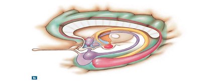
The Adult Human Brain
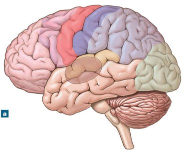
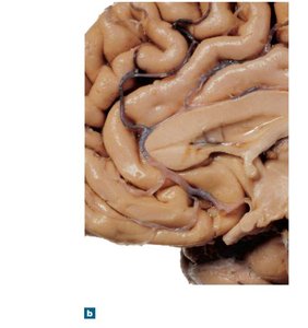
The adult human brain is the central organ of the nervous system, containing nearly 97% of the body's neural tissue. It ranges in volume from 750 to 2100 cc and weighs about 1.4 kg (3 lbs). The brain is responsible for processing sensory information, coordinating movement, and higher mental functions such as reasoning, memory, and emotion.
Cerebrum: Largest part, responsible for conscious thought, memory, and voluntary muscle activity.
Cerebellum: Coordinates repetitive body movements and maintains posture and balance.
Diencephalon: Contains thalamus and hypothalamus, relaying sensory information and regulating autonomic functions.
Brain Stem: Includes midbrain, pons, and medulla oblongata; controls vital functions and relays information between brain and spinal cord.
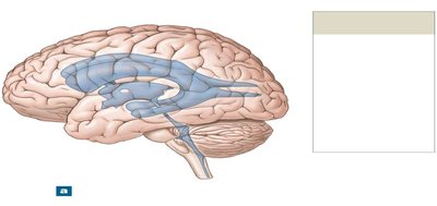
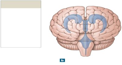
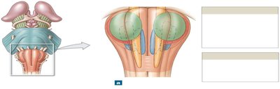
Ventricles of the Brain
Structure and Function
The brain contains a series of interconnected cavities called ventricles, which are filled with cerebrospinal fluid (CSF). These ventricles are remnants of the embryonic neural tube and are lined with ependymal cells. The main ventricles include:
Lateral Ventricles: One in each cerebral hemisphere, separated by the septum pellucidum.
Third Ventricle: Located in the diencephalon, connected to lateral ventricles via the interventricular foramen.
Fourth Ventricle: Located between the pons and cerebellum, continuous with the central canal of the spinal cord and connected to the third ventricle by the cerebral aqueduct.
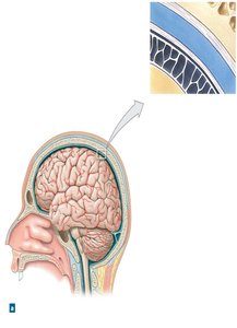
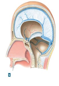
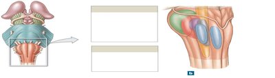
Protection and Support of the Brain
Cranial Meninges
The brain is protected by three connective tissue layers called meninges:
Dura Mater: Outermost, tough layer with two sublayers (endosteal and meningeal) and venous sinuses between them.
Arachnoid Mater: Middle, web-like layer with a subarachnoid space filled with CSF.
Pia Mater: Innermost, delicate layer attached to the brain surface by astrocytes.
Dural folds (falx cerebri, tentorium cerebelli, falx cerebelli) stabilize and support the brain within the cranial cavity.
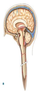
Cerebrospinal Fluid (CSF)
CSF surrounds all exposed surfaces of the CNS, cushioning neural structures, supporting the brain, and transporting nutrients, chemical messengers, and waste products. It is produced by the choroid plexus and circulates through the ventricles, central canal, and subarachnoid space. CSF is absorbed into the venous circulation via arachnoid granulations.
Blood Supply and Barriers
Blood–Brain Barrier (BBB): Formed by tight junctions between endothelial cells, isolating CNS tissue from general circulation and selectively allowing passage of substances.
Blood–CSF Barrier: Formed by special ependymal cells at the choroid plexus, allowing different chemical composition between blood and CSF.
Breaks in the BBB occur in regions such as the hypothalamus, posterior pituitary, pineal gland, and choroid plexus to allow hormone exchange.
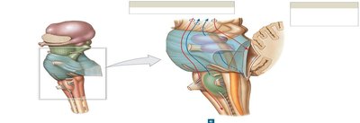
The Medulla Oblongata and Pons
Medulla Oblongata
The medulla oblongata connects the brain to the spinal cord and contains nuclei that regulate autonomic functions (cardiovascular and respiratory centers), relay sensory and motor information, and house cranial nerve nuclei.
Pons
The pons contains sensory and motor nuclei for cranial nerves, nuclei involved in respiratory control, and tracts that connect the cerebellum with the rest of the brain. It also contains transverse fibers linking the pons to the cerebellum.
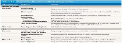
| Component | Function |
|---|---|---|
Medulla Oblongata | Nucleus gracilis, Nucleus cuneatus | Relay somatic sensory information to the thalamus |
Olivary nuclei | Relay information from the red nucleus, other nuclei of the midbrain, and the cerebral cortex to the cerebellum | |
Solitary nucleus | Integrates and relays visceral sensory information from the spinal and cranial nerves | |
Other sensory and motor nuclei | Relay sensory and motor information to higher centers | |
Reflex centers | Regulate heart rate and force of contraction | |
Respiratory rhythmicity centers | Set the pace of respiratory movements | |
Other autonomic centers | Regulate distribution of blood flow | |
Pons | Sensory and motor nuclei associated with cranial nerves V, VI, VII, and VIII | Relay sensory information and issue somatic motor commands |
Nuclei involved with the control of respiration | Modify output of respiratory centers in the medulla oblongata |
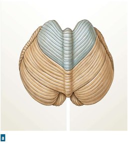
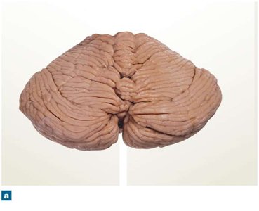
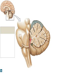
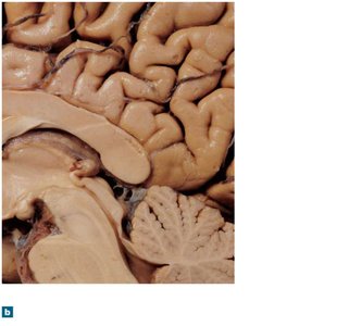
The Cerebellum
Structure and Function
The cerebellum coordinates voluntary movements, maintains posture, and ensures smooth, balanced muscular activity. It consists of two hemispheres, an anterior and posterior lobe, and is separated by the vermis. The cerebellar cortex is highly folded (folia), and the internal white matter is called the arbor vitae.
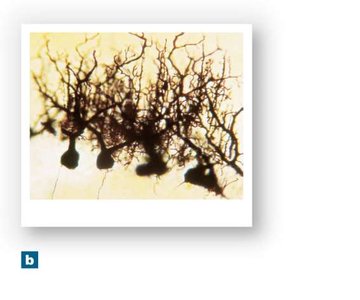
Purkinje cells: Large, branched neurons in the cerebellar cortex that receive extensive synaptic input.
Cerebellar peduncles: Tracts connecting the cerebellum to the brainstem (superior, middle, and inferior).
Subdivision | Region/Nuclei | Function |
|---|---|---|
Gray matter | Cerebellar cortex, Cerebellar nuclei | Involuntary coordination and control of ongoing body movements |
White matter | Arbor vitae | Connects cerebellar cortex and nuclei with cerebellar peduncles |
White matter | Cerebellar peduncles (superior, middle, inferior) | Link the cerebellum with midbrain, diencephalon, cerebrum, pons, and medulla oblongata |
White matter | Transverse fibers | Interconnect pontine nuclei with the cerebellar hemispheres on the opposite side |

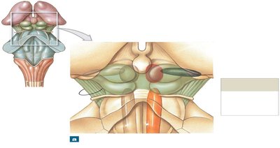
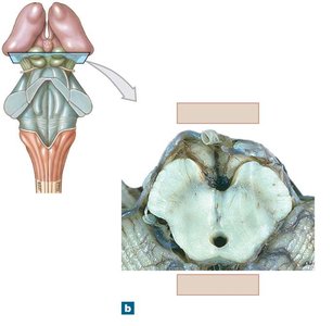
The Midbrain
Structure and Function
The midbrain (mesencephalon) is involved in processing visual and auditory information, maintaining consciousness, and controlling reflexive motor responses. Major structures include:
Tectum (Corpora quadrigemina): Superior colliculus (visual reflexes), Inferior colliculus (auditory reflexes)
Tegmentum: Red nucleus (motor coordination), Substantia nigra (regulates activity of basal nuclei)
Cerebral peduncles: Contain descending motor fibers and ascending sensory fibers
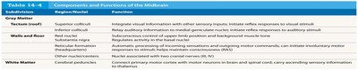
Gray Matter | Region/Nuclei | Function |
|---|---|---|
Tectum (roof) | Superior colliculi | Integrate visual information with other sensory inputs; initiate reflex responses to visual stimuli |
Tectum (roof) | Inferior colliculi | Relay auditory information to medial geniculate nucleus; initiate reflex responses to auditory stimuli |
Walls and floor | Red nucleus | Subconscious control of upper limb position and background muscle tone |
Walls and floor | Substantia nigra | Regulates activity in the basal nuclei |
Other | Reticular formation | Automatic processing of incoming sensations and outgoing motor commands; can initiate involuntary motor responses to stimuli; helps maintain consciousness |
White Matter | Cerebral peduncles | Carry descending motor commands from motor cortex to brain and spinal cord; carry ascending sensory information to thalamus |
The Diencephalon
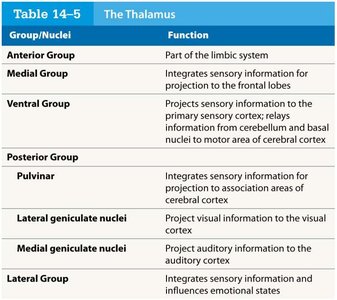
Thalamus
The thalamus acts as a relay and processing center for sensory information, filtering and directing signals to appropriate areas of the cerebral cortex. It consists of several nuclei, each with specific functions.
Group/Nuclei | Function |
|---|---|
Anterior Group | Part of the limbic system |
Medial Group | Integrates sensory information for projection to the frontal lobes |
Ventral Group | Projects sensory information to the primary sensory cortex; relays information from cerebellum and basal nuclei to motor area of cerebral cortex |
Posterior Group | Pulvinar: Integrates sensory information for projection to association areas of cerebral cortex |
Lateral geniculate nuclei | Project visual information to the visual cortex |
Medial geniculate nuclei | Project auditory information to the auditory cortex |
Lateral Group | Integrates sensory information and influences emotional states |
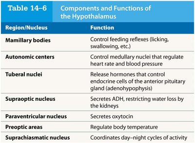
Hypothalamus
The hypothalamus is the primary center for autonomic control and endocrine integration. It regulates body temperature, hunger, thirst, circadian rhythms, and emotional responses. It also produces hormones such as ADH and oxytocin.
Region/Nucleus | Function |
|---|---|
Mamillary bodies | Control feeding reflexes (licking, swallowing, etc.) |
Autonomic centers | Control medullary nuclei that regulate heart rate and blood pressure |
Tuberal nuclei | Release hormones that control endocrine cells of the anterior pituitary gland |
Supraoptic nucleus | Secretes ADH, restricting water loss by the kidneys |
Paraventricular nucleus | Secretes oxytocin |
Preoptic areas | Regulate body temperature |
Suprachiasmatic nucleus | Coordinates day–night cycles of activity |
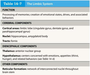
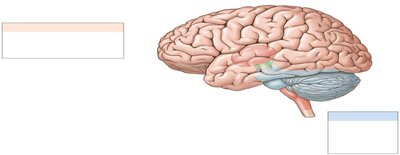
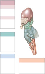
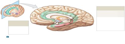
The Limbic System
Structure and Function
The limbic system is a functional grouping of structures involved in emotion, motivation, and memory. It links conscious intellectual functions with unconscious and autonomic functions.
Amygdaloid body: Interface between limbic system, cerebrum, and sensory systems
Limbic lobe: Includes cingulate, dentate, and parahippocampal gyri
Hippocampus: Essential for memory formation
Fornix: White matter tract connecting hippocampus with hypothalamus
Reticular formation: Influences emotional states
Function | Processing of memories; creation of emotional states, drives, and associated behaviors |
|---|---|
Cerebral Components | Limbic lobe (cingulate gyrus, dentate gyrus, parahippocampal gyrus), hippocampus, amygdaloid body, fornix |
Diencephalic Components | Thalamus (anterior nuclear group), hypothalamus (emotions, appetites, drives) |
Other Components | Reticular formation (network of nuclei throughout brain stem) |