 Back
BackThe Brain and Cranial Nerves: Structure, Development, and Function
Study Guide - Smart Notes
 Tailored notes based on your materials, expanded with key definitions, examples, and context.
Introduction to the Brain and Cranial Nerves
The adult human brain is the central organ of the nervous system, containing nearly 97% of the body's nervous tissue. It is responsible for processing sensory information, coordinating movement, and regulating vital functions. The brain is divided into several major regions, each with specialized roles in maintaining homeostasis and enabling complex behaviors.
Major Regions of the Brain
Cerebrum
The cerebrum is the largest part of the adult brain and is responsible for higher mental functions such as conscious thought, intellect, memory, and voluntary muscle control. It is divided into left and right hemispheres, each covered by a surface layer of gray matter called the cerebral cortex. The cortex is characterized by gyri (ridges), sulci (shallow grooves), and fissures (deep grooves), which increase its surface area.
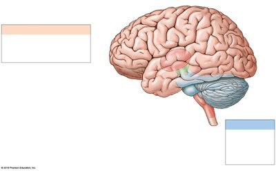
Cerebellum
The cerebellum is the second-largest brain region, coordinating repetitive body movements and maintaining posture and balance. It consists of two hemispheres and is covered by the cerebellar cortex (gray matter).
Diencephalon
The diencephalon lies beneath the cerebrum and cerebellum and includes the thalamus (sensory relay and processing), hypothalamus (emotions, autonomic function, hormone production), and the pituitary gland (major endocrine gland).
Brainstem
The brainstem connects the brain to the spinal cord and relays information between the cerebrum, cerebellum, and spinal cord. It consists of the midbrain, pons, and medulla oblongata, each with distinct functions in sensory and motor processing, reflexes, and autonomic regulation.
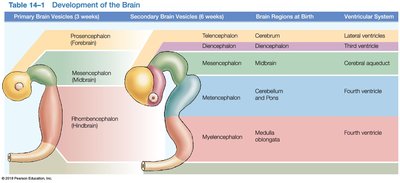
Development of the Brain
Embryonic Brain Vesicles
During embryonic development, the neural tube forms three primary brain vesicles: the prosencephalon (forebrain), mesencephalon (midbrain), and rhombencephalon (hindbrain). These further differentiate into secondary vesicles, which give rise to the major brain regions and ventricular system.
Primary Vesicle | Secondary Vesicle | Brain Region at Birth | Ventricular System |
|---|---|---|---|
Prosencephalon | Telencephalon | Cerebrum | Lateral ventricles |
Prosencephalon | Diencephalon | Diencephalon | Third ventricle |
Mesencephalon | Mesencephalon | Midbrain | Cerebral aqueduct |
Rhombencephalon | Metencephalon | Cerebellum and Pons | Fourth ventricle |
Rhombencephalon | Myelencephalon | Medulla oblongata | Fourth ventricle |
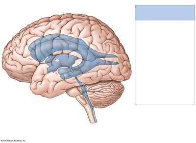
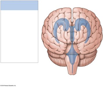
Ventricular System of the Brain
The brain contains a series of interconnected chambers called ventricles, which are filled with cerebrospinal fluid (CSF) and lined with ependymal cells. Each cerebral hemisphere contains a lateral ventricle, separated by the septum pellucidum. The third ventricle is located in the diencephalon, and the fourth ventricle extends into the medulla oblongata, connecting with the central canal of the spinal cord.
Protection and Support of the Brain
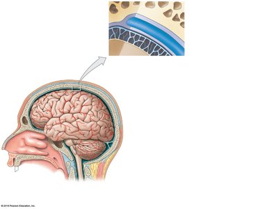
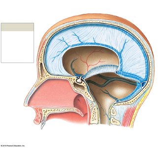
Cranial Meninges
The brain is protected by the bones of the cranium, three layers of cranial meninges (dura mater, arachnoid mater, pia mater), and cerebrospinal fluid. The dura mater forms dural folds that stabilize the brain and contain venous sinuses for blood drainage. The arachnoid mater covers the brain and is separated from the pia mater by the subarachnoid space, which contains CSF. The pia mater is attached to the brain surface by astrocytes.
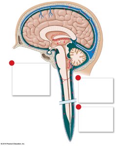
Cerebrospinal Fluid (CSF)
CSF surrounds all exposed surfaces of the CNS, supporting the brain, cushioning neural structures, and transporting nutrients, chemical messengers, and wastes. It is produced by the choroid plexus, circulates through the ventricles and subarachnoid space, and is absorbed into the venous circulation via arachnoid granulations.
Blood-Brain Barrier (BBB) and Blood-CSF Barrier
The BBB isolates the CNS from general circulation, formed by tight junctions between capillary endothelial cells and regulated by astrocytes. The blood-CSF barrier, formed by specialized ependymal cells, limits transfer of substances to CSF, allowing the chemical composition of blood and CSF to differ. Certain brain regions, such as the hypothalamus and choroid plexus, have breaks in the BBB to allow hormone release.
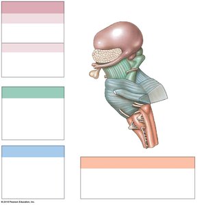
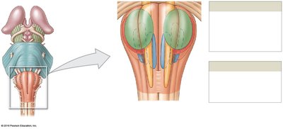
Brainstem: Medulla Oblongata, Pons, and Midbrain
Medulla Oblongata
The medulla oblongata is the most inferior part of the brainstem, coordinating complex autonomic reflexes and relaying sensory and motor information between the brain and spinal cord. It contains nuclei for cardiovascular and respiratory regulation, cranial nerves, and relay stations for sensory pathways.
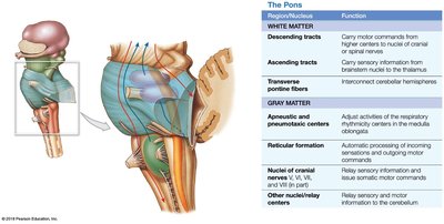
Pons
The pons contains sensory and motor nuclei for cranial nerves, nuclei involved in respiration, and tracts that relay information to and from the cerebellum. Transverse pontine fibers connect the pons with the cerebellar hemispheres.
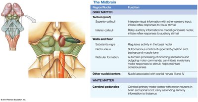
Midbrain
The midbrain (mesencephalon) contains the tectum (with superior and inferior colliculi for visual and auditory reflexes), tegmentum (red nucleus and substantia nigra), and cerebral peduncles (motor tracts). It is involved in processing sensory information, maintaining consciousness, and coordinating motor commands.
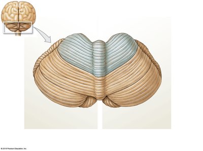
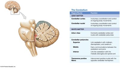
The Cerebellum
The cerebellum coordinates voluntary movements, maintains posture and balance, and fine-tunes motor activity. Its cortex is highly folded, and its internal white matter (arbor vitae) contains cerebellar nuclei. The cerebellum is connected to the brainstem by three pairs of cerebellar peduncles (superior, middle, inferior).
The Diencephalon
Thalamus
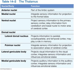
The thalamus acts as a relay and processing center for sensory information, filtering and transmitting signals to the cerebral cortex. It consists of several nuclei, each with specific functions in sensory integration, motor relay, and emotional processing.
Nuclei/Body | Function |
|---|---|
Anterior nuclei | Part of the limbic system |
Medial nuclei | Integrate sensory information for projection to the frontal lobes |
Ventral nuclei | Project sensory information to the primary sensory cortex; relay information from cerebellum and basal nuclei to motor area of cerebral cortex |
Lateral dorsal nucleus | Projects information to parietal, occipitoparietal, and temporal cortex; may play a role in memory |
Pulvinar nuclei | Integrate sensory information for projection to association areas of cerebral cortex |
Lateral geniculate body | Projects visual information to the visual cortex; integrates sensory information and influences emotional states |
Medial geniculate body | Projects auditory information to the auditory cortex; integrates sensory information and influences emotional states |
Hypothalamus
The hypothalamus regulates autonomic functions, hormone production, emotional responses, and circadian rhythms. It connects to the pituitary gland via the infundibulum and contains centers for hunger, thirst, and temperature regulation.
The Limbic System
The limbic system is a functional grouping of structures involved in emotion, motivation, and memory. It links conscious cortical functions with unconscious and autonomic processes, facilitating memory storage and retrieval.
The Cerebrum: Structure and Function
The cerebrum is responsible for conscious thought, intellectual functions, and processing somatic sensory and motor information. It is divided into lobes (frontal, parietal, temporal, occipital, insula) and contains gyri, sulci, and fissures that increase cortical surface area.
White Matter of the Cerebrum
Association fibers: Connect regions within the same hemisphere.
Commissural fibers: Connect the two hemispheres (e.g., corpus callosum).
Projection fibers: Link the cortex with lower brain regions and the spinal cord.
Basal Nuclei
The basal nuclei are masses of gray matter embedded in the white matter of the cerebrum. They direct subconscious activities, coordinate learned movement patterns, and regulate muscle tone.
Functional Areas of the Cerebral Cortex
Primary motor cortex: Controls voluntary movements (precentral gyrus).
Primary somatosensory cortex: Receives sensory information (postcentral gyrus).
Special sensory cortices: Visual, auditory, olfactory, and gustatory areas.
Association areas: Interpret sensory input and coordinate responses.
Integrative centers: Direct complex motor activities and analytical functions (e.g., Wernicke's and Broca's areas for language).
Cranial Nerves
There are 12 pairs of cranial nerves that emerge from the brain, each with specific sensory, motor, or mixed functions. They are essential for sensory perception, motor control, and autonomic regulation in the head, neck, and some visceral organs.
Olfactory (I): Smell
Optic (II): Vision
Oculomotor (III), Trochlear (IV), Abducens (VI): Eye movements
Trigeminal (V): Sensation in the face, motor to mastication muscles
Facial (VII): Facial expression, taste, glandular secretion
Vestibulocochlear (VIII): Hearing and balance
Glossopharyngeal (IX): Taste, swallowing, salivation
Vagus (X): Sensory and motor to thoracic and abdominal organs
Accessory (XI): Motor to neck and upper back muscles
Hypoglossal (XII): Motor to tongue muscles
Summary Table: Major Brain Regions and Functions
Region | Main Function(s) |
|---|---|
Cerebrum | Conscious thought, intellect, memory, voluntary movement |
Cerebellum | Coordination of movement, balance, posture |
Diencephalon | Sensory relay, autonomic and endocrine regulation |
Brainstem | Autonomic functions, reflexes, relay between brain and spinal cord |
