 Back
The Brain, Cranial Nerves, and Sensory and Motor Pathways: Structured Study Notes
Study Guide - Smart Notes
 Tailored notes based on your materials, expanded with key definitions, examples, and context.
The Brain, Cranial Nerves, and Sensory and Motor Pathways
Introduction
This chapter explores the structure, development, and function of the brain, cranial nerves, and the sensory and motor pathways. It covers the embryological origins of the brain, the major brain regions and their functions, protective structures, and the organization of sensory and motor systems.
Brain Development and Major Regions
Embryonic Development of the Brain
The brain develops from the embryonic neural tube, which forms three primary brain vesicles by the fourth week of development:
Prosencephalon (forebrain)
Mesencephalon (midbrain)
Rhombencephalon (hindbrain)
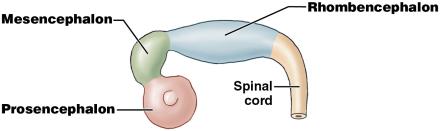
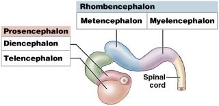
By the fifth week, these vesicles subdivide into secondary brain vesicles:
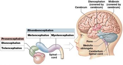
Prosencephalon → Telencephalon (forms cerebrum) and Diencephalon
Mesencephalon remains undivided
Rhombencephalon → Metencephalon (forms pons and cerebellum) and Myelencephalon (forms medulla oblongata)
Major Regions of the Adult Brain
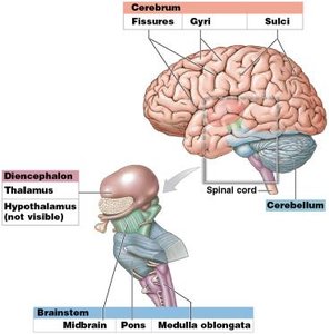
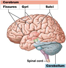
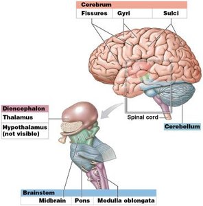
Cerebrum: Largest part, responsible for conscious thought, memory, and voluntary muscle activity.
Cerebellum: Coordinates and fine-tunes motor activity.
Diencephalon: Contains thalamus and hypothalamus; relays sensory information and controls autonomic functions.
Brainstem: Includes midbrain, pons, and medulla oblongata; controls vital autonomic functions and relays information.
Protection and Support of the Brain
Cranial Meninges
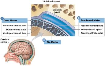
The brain is protected by three connective tissue layers called meninges:
Dura mater: Outermost, tough layer; has periosteal and meningeal layers.
Arachnoid mater: Middle, web-like layer; contains subarachnoid space filled with cerebrospinal fluid (CSF).
Pia mater: Innermost, delicate layer; adheres to brain surface.
Dural Folds and Sinuses
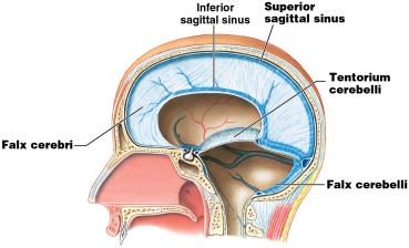
Dural folds stabilize the brain and contain large venous sinuses:
Falx cerebri: Between cerebral hemispheres
Tentorium cerebelli: Separates cerebrum from cerebellum
Falx cerebelli: Between cerebellar hemispheres
Cerebrospinal Fluid (CSF)
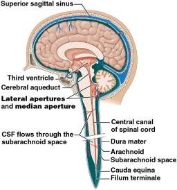
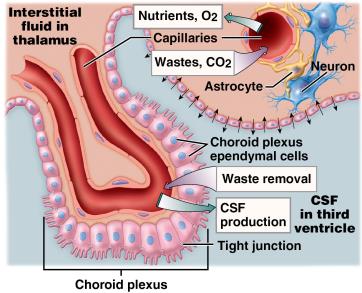
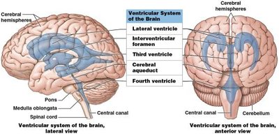
CSF supports, cushions, and nourishes the brain. It is produced by the choroid plexus in the ventricles and circulates through the ventricular system and subarachnoid space, eventually being absorbed into the venous circulation via arachnoid granulations.
Functions: Buoyancy, protection, chemical stability
Production: Choroid plexus (ependymal cells and capillaries)
Circulation: Lateral ventricles → third ventricle → cerebral aqueduct → fourth ventricle → subarachnoid space → arachnoid granulations
Functional Anatomy of Brain Regions
Cerebrum
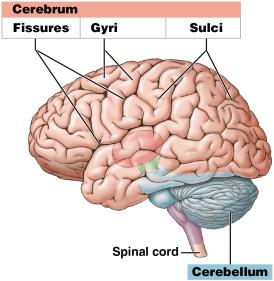
Divided into left and right hemispheres by longitudinal fissure
Surface features: Gyri (ridges), Sulci (grooves), Fissures (deep grooves)
Cerebral cortex: Thin outer layer of gray matter; site of conscious thought, memory, and sensory processing
Cerebellum
Coordinates and fine-tunes voluntary movements
Maintains posture and balance
Contains more than half of the brain's neurons
Diencephalon
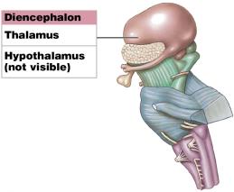
Thalamus: Relay and processing center for sensory information
Hypothalamus: Controls emotions, autonomic functions, and hormone production
Brainstem
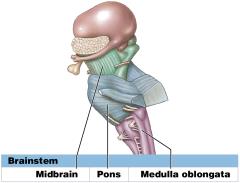
Midbrain: Processes visual and auditory information; controls reflexes
Pons: Connects cerebellum to brainstem; controls somatic and visceral motor functions
Medulla oblongata: Relays sensory information; regulates autonomic functions
Ventricular System of the Brain
The brain contains four interconnected ventricles filled with CSF:
Lateral ventricles: One in each cerebral hemisphere
Third ventricle: In the diencephalon
Fourth ventricle: Between pons and cerebellum
Summary Table: Major Brain Regions and Functions
Region | Main Structures | Primary Functions |
|---|---|---|
Cerebrum | Cerebral cortex, white matter, basal nuclei | Conscious thought, memory, voluntary movement |
Cerebellum | Cerebellar cortex, arbor vitae | Coordination of movement, balance |
Diencephalon | Thalamus, hypothalamus, epithalamus | Sensory relay, autonomic control, hormone production |
Brainstem | Midbrain, pons, medulla oblongata | Autonomic functions, relay between brain and spinal cord |
Key Terms and Concepts
Gyri: Elevated ridges on the cerebral surface
Sulci: Shallow grooves between gyri
Fissures: Deep grooves separating major brain regions
Choroid plexus: Network of capillaries producing CSF
Arachnoid granulations: Structures where CSF is absorbed into venous blood
Dural venous sinuses: Large veins within dural folds draining blood from the brain
Formulas and Equations
CSF Production Rate:
Additional Info
The brain's protection is a combination of physical barriers (skull, meninges), fluid cushioning (CSF), and biochemical isolation (blood-brain barrier).
Damage to specific brain regions can result in characteristic neurological deficits, emphasizing the importance of their specialized functions.
