Back
BackThe Brain: Diencephalon, Brainstem, Cerebellum, and Protective Structures
Study Guide - Smart Notes
 Tailored notes based on your materials, expanded with key definitions, examples, and context.
Diencephalon
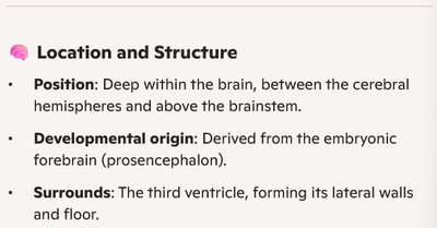
Location and Structure
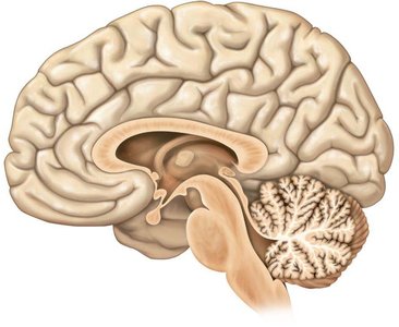
The diencephalon is a central brain region located deep within the brain, between the cerebral hemispheres and above the brainstem. It is derived from the embryonic forebrain (prosencephalon) and surrounds the third ventricle, forming its lateral walls and floor.
Overview and Functions
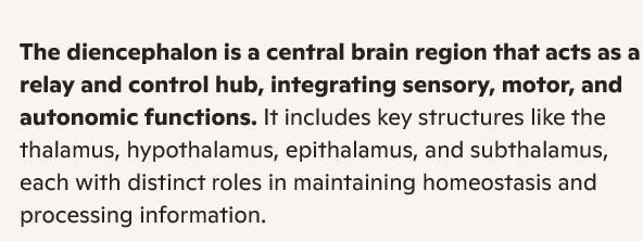
The diencephalon acts as a relay and control hub, integrating sensory, motor, and autonomic functions. It includes key structures such as the thalamus, hypothalamus, epithalamus, and subthalamus, each with distinct roles in maintaining homeostasis and processing information.
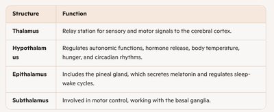
Major Structures of the Diencephalon
Structure | Function |
|---|---|
Thalamus | Relay station for sensory and motor signals to the cerebral cortex. |
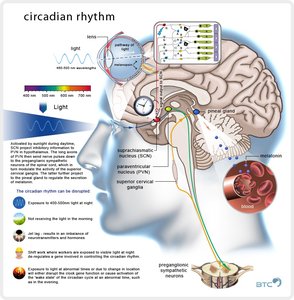
Hypothalamus | Regulates autonomic functions, hormone release, body temperature, hunger, and circadian rhythms. |
Epithalamus | Includes the pineal gland, which secretes melatonin and regulates sleep-wake cycles. |
Subthalamus | Involved in motor control, working with the basal ganglia. |
Key Anatomical Relationships
The diencephalon is closely associated with other brain regions, including the limbic system and the brainstem. It is positioned near the thalamus and hypothalamus, which are critical for sensory processing and homeostatic regulation.

Brainstem
Regions and Functions
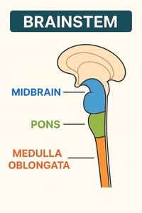
The brainstem is the lower part of the brain that connects to the spinal cord. It consists of three main regions:
Midbrain: Located just below the thalamus; controls visual and auditory reflexes, and is important for motor movement and coordination.
Pons: The middle section; acts as a bridge between the cerebrum and cerebellum, regulates breathing, and facilitates communication between brain regions.
Medulla Oblongata: The lowest part, continuous with the spinal cord; controls vital autonomic functions such as heart rate, blood pressure, and respiration.
Functions of the Brainstem:
Basic life support (breathing, heartbeat)
Reflexes (swallowing, coughing)
Pathway for sensory and motor signals between brain and body
Cerebellum
Location, Structure, and Functions
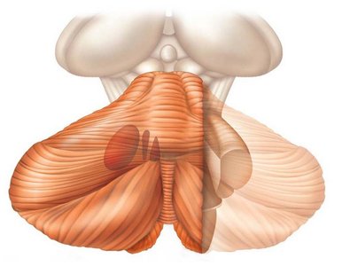
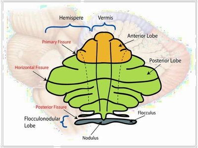
The cerebellum is located in the posterior part of the brain, beneath the occipital lobes and behind the brainstem. It consists of two hemispheres connected by the vermis, with a highly folded surface (folia). The cerebellum contains both grey matter (cerebellar cortex and deep nuclei) and white matter (arbor vitae and cerebellar peduncles).
Motor Coordination: Fine-tunes voluntary movements.
Balance & Posture: Maintains equilibrium.
Motor Learning: Important for learning new motor skills.
Cognitive Roles: Involved in attention and language processing.
Cerebellar Lobes and Structures
Anterior Lobe: Coordinates posture and limb movements.
Posterior Lobe: Fine motor control and voluntary movement.
Flocculonodular Lobe: Maintains balance and eye movements.
Vermis: Connects the two hemispheres and helps with posture control.
Functional Brain Systems
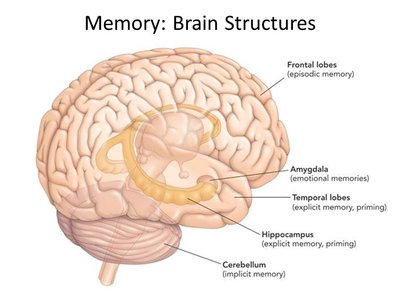
Limbic System
The limbic system is a group of interconnected structures involved in emotion, memory, and motivation. It includes:
Amygdala: Expression of emotion, arousal, and fear; associates stimuli with emotional value.
Hippocampus: Memory formation and learning.
Cingulate and Parahippocampal Gyri: Emotional processing and regulation.
Fornix: Main output tract of the limbic system.
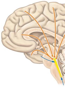
Reticular Formation
The reticular formation is a network of nuclei throughout the brainstem that receives input from multiple sources and sends output to the entire brain and spinal cord. It is involved in sleep, pain transmission, mood, motor functions, breathing, blood pressure, and alertness.
Higher Brain Functions
Language
Broca’s Area: Involved in speech production.
Wernicke’s Area: Involved in language comprehension.
Memory
Declarative Memory: Facts and events (short-term and long-term).
Procedural Memory: Skills and motor tasks.
Emotional Memory: Emotional responses to stimuli.
Factors influencing memory include emotional state, rehearsal, association, and automatic memory formation.
Brain Wave Patterns
Continuous electrical activity in the brain is measured by an electroencephalogram (EEG), which produces wave-like patterns:
Alpha (8-13 Hz): Calm wakefulness
Beta (14-30 Hz): Mental alertness and concentration
Theta (4-7 Hz): Common in children, appears during concentration
Delta (<5 Hz): High amplitude, observed during sleep
Sleep/Wake Cycles
Non-rapid eye movement (NREM): Four stages, from relaxation to deep sleep
Rapid eye movement (REM): Dreaming, muscle inhibition (except eyes and diaphragm)
Regulated by the circadian rhythm and hypothalamus; sleep is important for memory and emotional analysis.
Consciousness
Consciousness involves simultaneous activity of large areas of the cerebral cortex and is measured on a gradient from alertness to coma.
Protection of the Brain
Protective Structures
Skull Bones: Provide rigid protection.
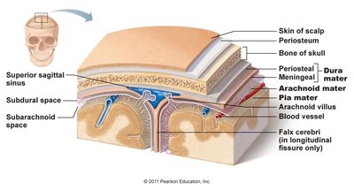
Cranial Meninges: Three protective membranes (dura mater, arachnoid mater, pia mater) of dense, irregular connective tissue.
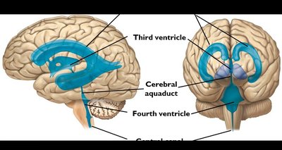
Cerebrospinal Fluid (CSF): Fluid with the same density as the brain, fills ventricles and surrounds the brain, providing buoyancy, temperature regulation, and waste removal.
Blood Brain Barrier (BBB): Separates CSF and brain extracellular fluid from blood, protecting the brain from toxins and pathogens.
Meninges
Dura Mater: Strongest, bi-layered sheet (periosteal and meningeal layers); forms dural venous sinuses and partitions (falx cerebri, falx cerebelli, tentorium cerebelli).
Arachnoid Mater: Middle meninx, separated from dura mater by the subarachnoid space filled with CSF.
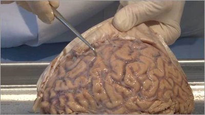
Pia Mater: Innermost layer, embedded with vasculature, closely adheres to the brain surface.
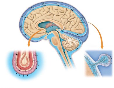
Cerebrospinal Fluid (CSF)
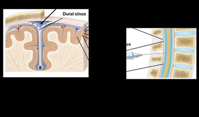
CSF is formed in the choroid plexuses of the ventricles, where blood vessels contact ependymal cells, extracting and converting plasma to CSF. CSF circulates through the ventricles, subarachnoid space, and central canal, and is reabsorbed into the bloodstream via dural sinuses (arachnoid granules). It provides buoyancy and protection, and is similar in composition to blood plasma but with more Na+, Cl-, and H+, and less Ca2+ and K+.
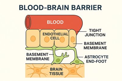
Blood Brain Barrier (BBB)
The BBB is a selective barrier that blocks toxins, pathogens, and large molecules from entering the brain, while allowing essential nutrients like glucose and amino acids to pass through. It maintains a stable environment for neurons. The main components include:
Component | Description |
|---|---|
Blood | The circulating fluid carrying nutrients, oxygen, and potential toxins. |
Endothelial Cells | Specialized cells lining brain capillaries; form the main barrier structure. |
Tight Junctions | Seal gaps between endothelial cells, preventing most substances from passing. |
Basement Membrane | Thin layer of extracellular matrix providing structural support to capillaries. |
Astrocyte End-Feet | Extensions of astrocytes that wrap around capillaries, aiding BBB maintenance. |
Brain Tissue | Neural tissue protected by the BBB from harmful substances in the blood. |