 Back
BackThe Brain: Structure, Function, and Associated Systems
Study Guide - Smart Notes
 Tailored notes based on your materials, expanded with key definitions, examples, and context.
Brain Structure and Organization
Major Regions of the Brain
The brain is divided into several major regions, each with distinct anatomical and functional characteristics. Understanding these regions is essential for grasping the complexity of neural processing and integration.
Cerebrum: The largest part, responsible for higher cognitive functions, voluntary movement, and sensory perception.
Diencephalon: Includes the thalamus, hypothalamus, and epithalamus; involved in sensory relay, autonomic control, and endocrine regulation.
Brainstem: Composed of the midbrain, pons, and medulla; controls vital autonomic functions and acts as a conduit for neural pathways.
Cerebellum: Coordinates movement, balance, and posture.
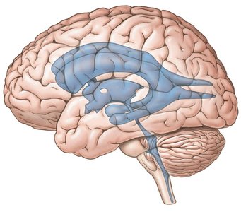
Cerebrospinal Fluid (CSF)
Functions and Circulation of CSF
Cerebrospinal fluid (CSF) is a clear, colorless liquid that surrounds the brain and spinal cord, providing mechanical and chemical protection, as well as facilitating nutrient and waste exchange.
Mechanical Protection: Acts as a shock absorber for the brain and spinal cord.
Chemical Protection: Maintains an optimal environment for neuronal signaling.
Circulation: Enables exchange of nutrients and waste between blood and nervous tissue.
CSF is produced by the choroid plexus in the ventricles and is reabsorbed by the arachnoid villi into the superior sagittal sinus, maintaining constant pressure.
Ventricles: Lateral ventricles (one in each hemisphere), third ventricle (between thalamic halves), and fourth ventricle (between brainstem and cerebellum).
Flow Pathway: Lateral ventricles → interventricular foramen → third ventricle → cerebral aqueduct → fourth ventricle → central canal/subarachnoid space → reabsorption via arachnoid villi.
Cerebrum
Anatomy and Functional Areas
The cerebrum is divided into two hemispheres and four main lobes, each associated with specific functions. The surface is highly folded, increasing surface area for cortical processing.
Gyri: Outward folds of the cortex.
Sulci: Shallow grooves between gyri.
Fissures: Deep grooves, such as the longitudinal fissure (separates hemispheres) and transverse fissure (separates cerebrum from cerebellum).
Cerebral Cortex: Superficial gray matter responsible for conscious thought and voluntary actions.
Cerebral White Matter: Contains myelinated tracts for communication between regions.
Corpus Callosum: Major white matter tract connecting the hemispheres, enabling interhemispheric communication.
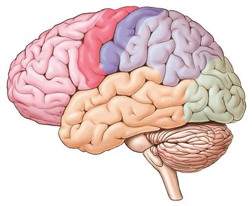
Lobes of the Cerebrum and Their Functions
Frontal Lobe: Personality, planning, voluntary movement, speech production (Broca’s area).
Parietal Lobe: Processes sensory information from the body (primary sensory cortex in postcentral gyrus).
Occipital Lobe: Visual processing and memory storage.
Temporal Lobe: Auditory, olfactory, and taste perception.
Cerebral lateralization refers to the specialization of each hemisphere for certain tasks (e.g., language in the left, spatial abilities in the right).
Cerebral Nuclei (Basal Ganglia)
The basal ganglia are deep gray matter structures that regulate motor output and inhibit unwanted movements. They include the caudate nucleus, lentiform nucleus, claustrum, and amygdaloid body.
Functional Systems
Limbic System: Governs emotions and memory.
Reticular Formation: Maintains consciousness and alertness.
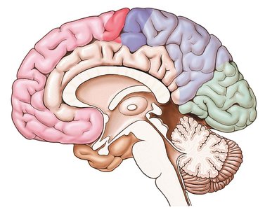
Diencephalon
Components and Functions
The diencephalon is located between the cerebral hemispheres and the brainstem, surrounding the third ventricle.
Thalamus: Relay station for sensory information (except smell) to the cerebral cortex.
Hypothalamus: Regulates autonomic functions, body temperature, hunger, thirst, and controls the endocrine system.
Epithalamus: Contains the pineal gland (produces melatonin) and habenular nuclei (emotional response to odors).
Brainstem
Structure and Function
The brainstem connects the cerebrum with the spinal cord and is essential for basic life functions.
Midbrain: Contains the substantia nigra (dopamine production), red nucleus (posture), and tectum (visual and auditory reflex centers).
Pons: Regulates breathing and assists in sound localization.
Medulla Oblongata: Contains centers for cardiovascular and respiratory control, as well as reflexes for coughing, swallowing, and vomiting.
Cerebellum
Role in Motor Control
The cerebellum is responsible for coordinating voluntary movements, maintaining muscle tone, and ensuring balance and posture. It receives input from sensory systems and fine-tunes motor activity.
Cranial Nerves
Overview and Functions
There are twelve pairs of cranial nerves, each with specific sensory, motor, or mixed functions. They innervate structures primarily in the head and neck.
Number | Name | Type | Main Function(s) |
|---|---|---|---|
CN I | Olfactory | Sensory | Smell |
CN II | Optic | Sensory | Vision |
CN III | Oculomotor | Motor/Parasympathetic | Eye movement, pupil constriction |
CN IV | Trochlear | Motor | Eye movement (SO muscle) |
CN V | Trigeminal | Mixed | Facial sensation, mastication |
CN VI | Abducens | Motor | Eye movement (LR muscle) |
CN VII | Facial | Mixed/Parasympathetic | Facial expression, taste (anterior 2/3 tongue), glands |
CN VIII | Vestibulocochlear | Sensory | Hearing, balance |
CN IX | Glossopharyngeal | Mixed/Parasympathetic | Taste (posterior 1/3 tongue), swallowing, parotid gland |
CN X | Vagus | Mixed/Parasympathetic | Viscera, swallowing, speech |
CN XI | Spinal Accessory | Motor | Trapezius, SCM muscles |
CN XII | Hypoglossal | Motor | Tongue movement |
Autonomic Nervous System (ANS) Responses
Sympathetic and Parasympathetic Divisions
The autonomic nervous system (ANS) regulates involuntary physiological functions and maintains homeostasis through dual innervation of most organs.
Sympathetic Division: Prepares the body for 'fight-or-flight' responses (emergency, exercise, excitement, embarrassment). Effects include increased heart rate, blood pressure, and energy mobilization.
Parasympathetic Division: Promotes 'rest-and-digest' activities (salivation, digestion, urination, defecation, decreased heart rate and pupil diameter).
Autonomic Tone: The balance between sympathetic and parasympathetic activity, allowing fine control of organ function.
Example: During exercise, sympathetic activity increases heart rate and redirects blood flow to muscles, while parasympathetic activity predominates during rest, promoting digestion and energy conservation.
