 Back
BackThe Brain: Structure, Function, and Associated Systems
Study Guide - Smart Notes
 Tailored notes based on your materials, expanded with key definitions, examples, and context.
Brain Structure and Organization
Major Regions of the Brain
The brain is divided into several major regions, each with distinct anatomical and functional characteristics. Understanding these regions is essential for comprehending higher-order functions, sensory processing, and motor control.
Cerebrum: The largest part, responsible for higher functions such as thought, memory, sensation, and voluntary movement. It consists of two hemispheres and four lobes, with a highly folded surface of gyri (ridges) and sulci (grooves).
Diencephalon: Located centrally, includes the thalamus, hypothalamus, and epithalamus. It surrounds the third ventricle and is involved in sensory relay, autonomic, and endocrine functions.
Brainstem: Composed of the midbrain, pons, and medulla oblongata. It controls vital autonomic functions and acts as a conduit for information between the brain and spinal cord.
Cerebellum: Located posteriorly, it coordinates voluntary movements, balance, and posture.
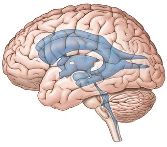
Cerebrospinal Fluid (CSF)
Functions and Circulation of CSF
Cerebrospinal fluid (CSF) is a clear, colorless liquid that nourishes and protects the brain and spinal cord. It circulates through the subarachnoid space, brain ventricles, and central canal of the spinal cord.
Mechanical Protection: Acts as a shock absorber for the brain and spinal cord.
Chemical Protection: Maintains an optimal environment for neuronal signaling.
Circulation: Facilitates exchange of nutrients and waste between blood and nervous tissue.
CSF is produced by the choroid plexus (capillary network covered by ependymal cells) in the ventricles and is replaced every 3-4 hours. Its composition includes glucose, proteins, ions, and some white blood cells.
Ventricles of the Brain
Lateral Ventricles: One in each cerebral hemisphere.
Third Ventricle: Located between the halves of the thalamus.
Fourth Ventricle: Between the brainstem and cerebellum.
CSF Circulation Pathway
CSF flows from the lateral ventricles through the interventricular foramen into the third ventricle.
From the third ventricle, it enters the cerebral aqueduct and then the fourth ventricle.
CSF exits to the central canal or subarachnoid space via median and lateral apertures.
It circulates in the subarachnoid space and is reabsorbed into the superior sagittal sinus by arachnoid villi, maintaining constant pressure.
Cerebrum: Structure and Functional Areas
Anatomy of the Cerebrum
The cerebrum is divided into two hemispheres, each with four lobes. The surface is marked by gyri (ridges) and sulci (grooves), which increase surface area for cortical processing.
Longitudinal Fissure: Separates the two hemispheres.
Transverse Fissure: Divides the cerebrum from the cerebellum.
Central Sulcus: Separates the frontal and parietal lobes.
Corpus Callosum: A white matter tract connecting the hemispheres, enabling communication.
Cerebral Lateralization
Right Hemisphere: Controls the left side of the body; involved in spatial, artistic, and emotional processing.
Left Hemisphere: Controls the right side; responsible for reasoning, language, and scientific skills.
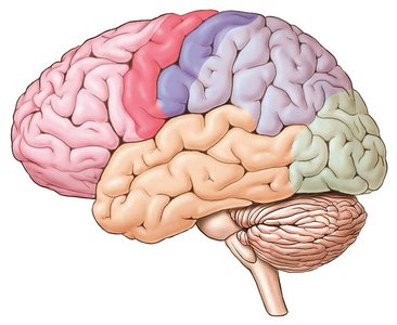
Lobes of the Cerebrum and Their Functions
Frontal Lobe: Personality, planning, voluntary movement, and speech (Broca’s area).
Parietal Lobe: Processes sensory information (primary sensory cortex in the postcentral gyrus).
Occipital Lobe: Visual processing and memory storage.
Temporal Lobe: Hearing, smell, and taste perception.
Cerebral Nuclei (Basal Ganglia)
Includes the caudate nucleus, lentiform nucleus, claustrum, and amygdaloid body.
Regulates motor output and inhibits unwanted movements.
Functional Areas
Limbic System: Responsible for emotions.
Reticular Formation: Maintains consciousness.
Diencephalon
Regions and Functions
Thalamus: Relay station for sensory signals (except smell) to the cerebrum.
Hypothalamus: Regulates body temperature, hunger, thirst, autonomic and endocrine functions.
Epithalamus: Contains the pineal gland (produces melatonin) and habenular nucleus (emotional response to odors).
Brainstem
Components and Functions
Midbrain: Contains the substantia nigra (dopamine production), red nucleus (posture), and tectum (visual and auditory reflex centers).
Pons: Regulates breathing transitions and helps localize sound.
Medulla Oblongata: Contains motor tracts, cardiovascular and respiratory centers, and nuclei for reflexes such as coughing and swallowing.
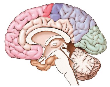
Cerebellum
Role in Motor Control
The cerebellum is responsible for coordinating skeletal muscle movements, maintaining muscle tone, and ensuring balance and posture. It receives proprioceptive input and fine-tunes motor activity.
Cranial Nerves
Overview and Functions
There are twelve pairs of cranial nerves, each with specific sensory, motor, or mixed functions. They innervate structures primarily in the head and neck.
Number | Name | Type | Main Function(s) |
|---|---|---|---|
CN I | Olfactory | Sensory | Smell |
CN II | Optic | Sensory | Vision |
CN III | Oculomotor | Motor/Parasympathetic | Eye movement, pupil diameter, lens adjustment |
CN IV | Trochlear | Motor | Eye movement (SO muscle) |
CN V | Trigeminal | Mixed | Mastication, facial sensation |
CN VI | Abducens | Motor | Eye movement (LR muscle) |
CN VII | Facial | Mixed/Parasympathetic | Facial expression, taste (anterior 2/3), glands |
CN VIII | Vestibulocochlear | Sensory | Balance, hearing |
CN IX | Glossopharyngeal | Mixed/Parasympathetic | Swallowing, taste (posterior 1/3), parotid gland |
CN X | Vagus | Mixed/Parasympathetic | Swallowing, larynx, thoracic/abdominal organs |
CN XI | Spinal Accessory | Motor | Trapezius, SCM muscles |
CN XII | Hypoglossal | Motor | Tongue muscles |
Autonomic Nervous System (ANS) Responses
Sympathetic and Parasympathetic Divisions
The ANS regulates involuntary body functions and maintains homeostasis through dual innervation of most organs. The sympathetic and parasympathetic divisions generally have opposing effects.
Sympathetic Division ("Fight-or-Flight")
Prepares the body for emergencies (stimulated by Emergency, Embarrassment, Exercise, Excitement).
Effects: Pupil dilation, increased heart rate and blood pressure, decreased blood flow to kidneys/GI tract, increased blood flow to muscles, liver, heart, and adipose tissue, glucose release, fat breakdown.
Effects are longer-lasting and more widespread than parasympathetic responses.
Parasympathetic Division ("Rest-and-Digest")
Promotes energy conservation and restoration during rest.
Effects: Salivation, lacrimation, urination, digestion, defecation (SLUDD), decreased heart rate, airway diameter, and pupil diameter.
Effects are shorter and less widespread than sympathetic responses.
Summary Table: Brain Regions and Major Functions
Region | Main Functions |
|---|---|
Cerebrum | Higher cognitive functions, voluntary movement, sensory processing |
Diencephalon | Sensory relay, autonomic and endocrine regulation |
Brainstem | Autonomic control, reflexes, conduit for information |
Cerebellum | Coordination, balance, posture |
