 Back
BackThe Brain: Structure, Function, and Major Pathways (Chapter 14 Study Notes)
Study Guide - Smart Notes
 Tailored notes based on your materials, expanded with key definitions, examples, and context.
Brain Structure and Major Divisions
Cerebrum
The cerebrum is the largest part of the brain and is responsible for higher cognitive functions such as reasoning, planning, and voluntary movement. It is divided into two cerebral hemispheres, each containing folds called gyri and grooves called sulci.
Gyri: Elevated ridges on the brain's surface.
Sulci: Shallow grooves separating gyri.
Longitudinal fissure: Deep groove separating right and left hemispheres.
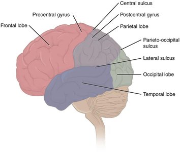
Major Lobes and Functions:
Frontal lobe: Voluntary motor functions, judgment, planning, memory, taste.
Parietal lobe: Sensory input processing.
Occipital lobe: Visual processing center.
Temporal lobe: Hearing, smell, learning, memory, visual recognition.
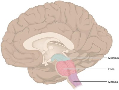
Brain Stem
The brain stem connects the cerebrum with the spinal cord and is essential for vital functions such as breathing and heart rate. It consists of the midbrain, pons, and medulla oblongata.
Midbrain: Visual and auditory reflexes, dopamine production.
Pons: Respiratory control, bridge for neural pathways.
Medulla oblongata: Cardiac, respiratory, and vasomotor centers; decussation of motor tracts.
Cerebellum
The cerebellum is located posterior to the brain stem and is responsible for balance, equilibrium, and coordination of voluntary movements. It compares intended movements with actual movements and makes adjustments as needed.
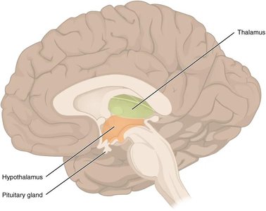
Diencephalon
The diencephalon is situated deep within the cerebrum and includes the thalamus, hypothalamus, and epithalamus. It is a major center for homeostasis and sensory/motor integration.
Thalamus: Relay station for sensory and motor signals to the cortex.
Hypothalamus: Regulates appetite, thirst, body temperature, autonomic nervous system, and hormone production.
Epithalamus: Contains the pineal gland, which produces melatonin for sleep-wake cycles.
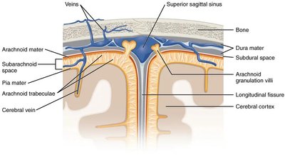
Meninges and Protective Coverings
Meningeal Layers
The meninges are three connective tissue membranes that surround the brain and spinal cord, providing protection and support.
Dura mater: Tough, outermost layer; periosteal layer fused to skull, meningeal layer extends into fissures.
Arachnoid mater: Middle, web-like layer; closely adheres to dura.
Pia mater: Thin, innermost layer; follows contours of the brain into sulci.
The subarachnoid space between the arachnoid and pia mater contains cerebrospinal fluid (CSF).
Hematomas
Epidural hematoma: Blood collects between dura and skull, often due to trauma; increases intracranial pressure.
Subdural hematoma: Blood collects between dura and arachnoid; also increases pressure and can cause brain damage.
Cerebrospinal Fluid (CSF)
Functions of CSF
Cerebrospinal fluid is a clear, colorless liquid that surrounds the brain and spinal cord, providing several essential functions:
Maintains a stable environment for neural tissue
Provides nutrition and removes waste
Protects the brain by cushioning against trauma
Regulates intracranial pressure
Composition of CSF
99% water
Ions: Na+, Cl-, Mg++
Much less protein than blood
Glucose (about 70% of blood level)
Very few cells
CSF Circulation Pathway
CSF is produced by the choroid plexus in the lateral ventricles and flows through the ventricular system:
Lateral ventricles
Interventricular foramen
Third ventricle
Cerebral aqueduct
Fourth ventricle
Subarachnoid space (or central canal)
Arachnoid villi
Superior sagittal sinus (venous return)
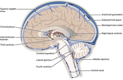
Clinical Note: Hydrocephalus
Hydrocephalus occurs when more CSF is produced than reabsorbed, leading to water accumulation in the brain. This condition can cause increased intracranial pressure and requires medical intervention.
Functional Organization of the Cerebrum
Gray and White Matter
Gray matter (cortex): Contains neuron cell bodies; site of major brain functions.
White matter (tracts): Contains myelinated axons; connects different brain regions.
Types of White Matter Tracts
Commissural tracts: Connect right and left hemispheres (e.g., corpus callosum).
Association tracts: Connect gyri and lobes within the same hemisphere.
Projection tracts: Connect cortex with lower brain regions and spinal cord.
Functional Areas of the Cortex
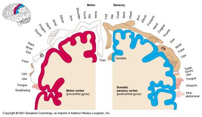
Motor cortex (precentral gyrus, frontal lobe): Initiates voluntary movements.
Sensory cortex (postcentral gyrus, parietal lobe): Receives sensory input.
Broca's area (frontal lobe): Motor speech production.
Wernicke's area (parietal lobe): Language comprehension and formulation.
Visual center (occipital lobe): Processes visual information.
Auditory center (temporal lobe): Processes auditory information.
Gustatory center (frontal lobe/insula): Processes taste.
Hemispheric Specialization
Right hemisphere: Controls left side of body; associated with music, artistic skills, spatial relationships, and insight.
Left hemisphere: Controls right side of body; associated with speech, numerical and reasoning skills.
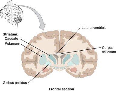
Basal Nuclei
Structure and Function
The basal nuclei are masses of gray matter deep within the white matter of the cerebrum. They are involved in the control of subconscious motor impulses, muscle tone, and coordination of movement.
Receive and send signals to the substantia nigra (midbrain) and cerebral cortex.
Work with the cerebellum and precentral gyrus to refine movements.
Dopamine from the substantia nigra inhibits excessive movement; loss of these cells leads to Parkinson's disease.
Diencephalon: Thalamus, Hypothalamus, and Epithalamus
Thalamus
Relay station for almost all sensory and motor information to the cortex.
Sorts, edits, and relays impulses by function.
Mediates sensation, motor activities, arousal, learning, and memory.
Hypothalamus
Regulates appetite, thirst, body temperature, and autonomic nervous system.
Controls the pituitary gland and hormone production.
Involved in emotions, memory, and circadian rhythms (with pineal gland).
Epithalamus
Contains the pineal gland, which produces melatonin to regulate sleep-wake cycles.
Works with the hypothalamus to control biorhythms.
Limbic System
The limbic system includes parts of the cerebral hemispheres and diencephalon. It is essential for emotions, memory, and motivation.
Brain Stem: Mesencephalon, Pons, and Medulla Oblongata
Mesencephalon (Midbrain)
Corpora quadrigemina: Superior colliculi (visual reflexes), inferior colliculi (auditory reflexes).
Tegmentum: Substantia nigra (dopamine), red nucleus (motor coordination).
Reticular formation: Controls alertness, sleep, and pain modulation.
Cerebral peduncles: Descending motor tracts.
Pons
Respiratory center.
Bridge for neural pathways between brain regions and cerebellum.
Medulla Oblongata
Contains vital centers for respiration, cardiac function, and vasomotor control.
Non-vital centers for vomiting, sneezing, and coughing.
Site of decussation (crossing) of motor tracts.
Cerebellum
The cerebellum coordinates voluntary movements, balance, and posture. It compares intended movements with actual performance and makes necessary adjustments. It works ipsilaterally (right side controls right body, left controls left).
Cranial Nerves
Overview
There are 12 pairs of cranial nerves, each with specific sensory, motor, or mixed functions. They emerge from the brain and brain stem and innervate structures primarily in the head and neck.
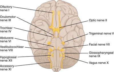
Number | Name | Type | Main Function(s) |
|---|---|---|---|
I | Olfactory | Sensory | Smell |
II | Optic | Sensory | Vision |
III | Oculomotor | Motor | Eye movement, pupil constriction |
IV | Trochlear | Motor | Eye movement (superior oblique muscle) |
V | Trigeminal | Both | Sensory to face, motor to mastication |
VI | Abducens | Motor | Eye movement (lateral rectus muscle) |
VII | Facial | Both | Taste (anterior 2/3 tongue), facial expression, tear/saliva secretion |
VIII | Vestibulocochlear | Sensory | Hearing, balance |
IX | Glossopharyngeal | Both | Taste (posterior 1/3 tongue), swallowing, salivation |
X | Vagus | Both | Viscera control, taste, swallowing, parasympathetic output |
XI | Accessory | Motor | Neck and shoulder movement, swallowing |
XII | Hypoglossal | Motor | Tongue movement |
Summary Table: Major Brain Regions and Functions
Region | Main Functions |
|---|---|
Cerebrum | Higher thinking, voluntary movement, sensory perception |
Brain Stem | Vital functions, cranial nerve origin, pathway for impulses |
Cerebellum | Balance, coordination, motor learning |
Diencephalon | Homeostasis, sensory/motor integration, hormone regulation |
