 Back
BackThe Brain: Structure, Protection, and Functional Organization
Study Guide - Smart Notes
 Tailored notes based on your materials, expanded with key definitions, examples, and context.
Major Regions and Overview of the Brain
Introduction to Brain Anatomy
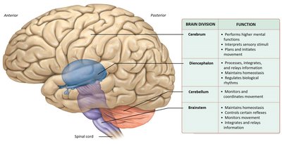
The brain is the central organ of the nervous system, responsible for processing sensory information, coordinating movement, and regulating vital functions. It is divided into several major regions, each with specialized roles in cognition, sensation, and homeostasis.
Cerebrum: Responsible for higher mental functions, including reasoning, memory, and voluntary movement.
Diencephalon: Integrates sensory information and maintains homeostasis.
Cerebellum: Coordinates movement and balance.
Brainstem: Controls basic life-sustaining functions such as breathing and heart rate.
Gray and White Matter Distribution
Structural Organization
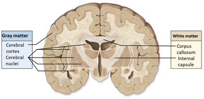
The brain consists of two main types of tissue: gray matter and white matter. Their distribution is essential for understanding brain function and connectivity.
Gray Matter: Contains neuron cell bodies, dendrites, and unmyelinated axons. Found in the outer cerebral cortex and deep cerebral nuclei (basal nuclei).
White Matter: Composed of myelinated axons organized into tracts, facilitating communication between different brain regions.
Protection and Support of the Brain
Protective Structures
The brain is protected by several anatomical structures that provide physical support and maintain a stable environment.
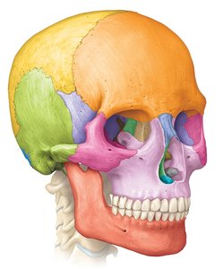
Bones of the Skull: Form a rigid protective case around the brain.
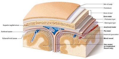
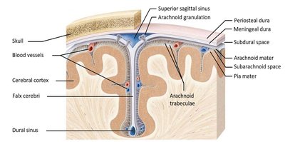
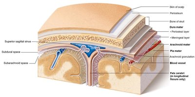
Cranial Meninges: Three connective tissue layers (dura mater, arachnoid mater, pia mater) that cover and protect the brain and spinal cord, contain cerebrospinal fluid (CSF), and enclose blood vessels.
Cerebrospinal Fluid (CSF): Cushions the brain, provides buoyancy, and transports nutrients and waste.
Blood-Brain Barrier: Regulates the movement of substances between the blood and brain tissue.
Cranial Meninges and Spaces
The meninges consist of three layers and associated spaces that play critical roles in brain protection and function.
Dura Mater: Tough, outermost membrane with two layers in the brain (periosteal and meningeal).
Arachnoid Mater: Thin, web-like middle layer containing arachnoid granulations for CSF reabsorption.
Pia Mater: Delicate inner layer bound to the brain surface by astrocytes.
Spaces: Subdural space (between dura and arachnoid), subarachnoid space (between arachnoid and pia, contains CSF).
Dural Folds
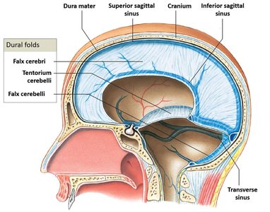
Dural folds are extensions of the dura mater that partition the cranial cavity and help stabilize the brain.
Falx Cerebri: Located in the longitudinal fissure, separates the two cerebral hemispheres.
Falx Cerebelli: Separates the two cerebellar hemispheres.
Tentorium Cerebelli: Separates the cerebrum from the cerebellum.
Ventricular System and Cerebrospinal Fluid (CSF)
Brain Ventricles
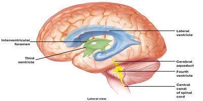
The ventricular system consists of interconnected cavities within the brain that produce, circulate, and contain CSF.
Lateral Ventricles: One in each cerebral hemisphere.
Third Ventricle: Located in the diencephalon, connected to lateral ventricles via interventricular foramen.
Fourth Ventricle: Located between the pons and cerebellum, connected to the third ventricle by the cerebral aqueduct.
CSF Production and Circulation
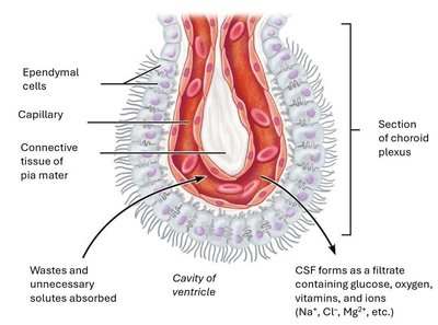
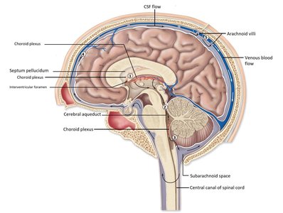
CSF is produced by the choroid plexus in the ventricles and circulates through the ventricular system and subarachnoid space before being reabsorbed into the venous blood.
Formation: Choroid plexus filters blood plasma to produce CSF.
Circulation: CSF flows from lateral ventricles → interventricular foramen → third ventricle → cerebral aqueduct → fourth ventricle → subarachnoid space and central canal.
Reabsorption: Occurs at arachnoid granulations into the superior sagittal sinus.
Functions of CSF
Buoyancy: Reduces the effective weight of the brain by 95%.
Protection: Cushions the brain against trauma.
Transport: Delivers nutrients and removes waste products.
Homeostatic Regulation: Maintains chemical stability and responds to changes detected by chemoreceptors.
Clinical Note: Disruption of CSF flow can lead to hydrocephalus, a condition characterized by abnormal accumulation of CSF in the ventricles.
Blood-Brain Barrier
Structure and Function
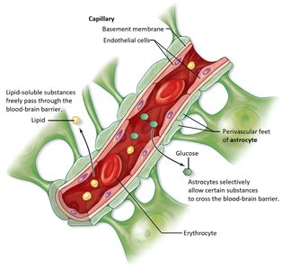
The blood-brain barrier (BBB) is a selective barrier formed by astrocytes and endothelial cells of brain capillaries. It regulates the passage of substances from the blood into the brain, protecting neural tissue from toxins and pathogens while allowing essential nutrients to pass.
Astrocytes: Wrap around capillaries and induce tight junctions between endothelial cells.
Selective Permeability: Allows passage of lipid-soluble substances, glucose, and some ions; restricts large or harmful molecules.
Cerebrum: Structure and Functional Areas
Gross Anatomy of the Cerebrum
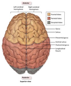
The cerebrum is the largest part of the brain, divided into two hemispheres and four main lobes. It is responsible for higher cognitive functions, voluntary movement, and sensory perception.
Lobes: Frontal, Parietal, Occipital, Temporal
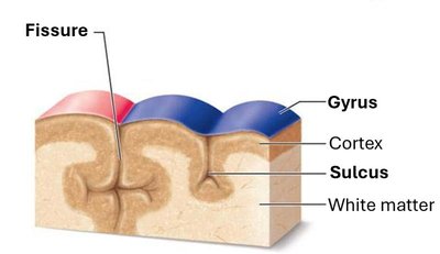
Surface Features: Gyri (ridges), sulci (shallow grooves), and fissures (deep grooves)
Functional Areas of the Cerebral Cortex
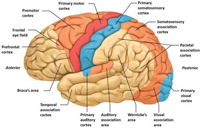
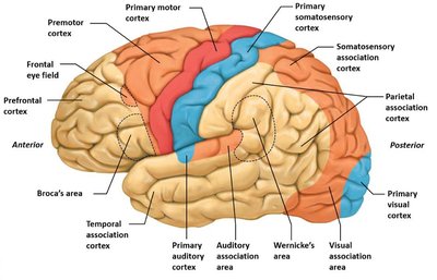
The cerebral cortex is organized into distinct functional areas responsible for processing sensory input, generating motor output, and integrating information.
Primary Motor Cortex: Located in the precentral gyrus; initiates voluntary movements.
Premotor Cortex: Plans and coordinates complex movements.
Primary Somatosensory Cortex: Located in the postcentral gyrus; receives sensory information from the body.
Association Areas: Integrate and interpret sensory and motor information.
Motor and Sensory Homunculi
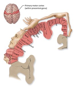
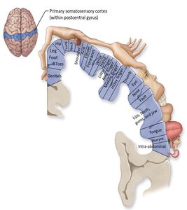
The motor and sensory homunculi are visual representations of the body regions controlled or sensed by specific areas of the cortex. The size of each region reflects the degree of innervation or sensitivity.
Motor Homunculus: Represents the distribution of motor control in the precentral gyrus.
Sensory Homunculus: Represents the distribution of sensory input in the postcentral gyrus.
Special Sensory and Association Areas
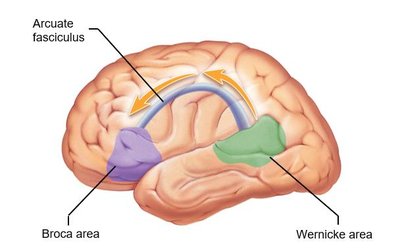
Specialized regions of the cortex process auditory, visual, olfactory, gustatory, and vestibular information. Language areas such as Broca's and Wernicke's areas are critical for speech production and comprehension.
Primary Auditory Cortex: Temporal lobe; processes sound.
Primary Visual Cortex: Occipital lobe; processes visual information.
Primary Olfactory Cortex: Temporal lobe; processes smell.
Primary Gustatory Cortex: Processes taste.
Language Areas: Broca's area (motor speech), Wernicke's area (sensory speech), connected by the arcuate fasciculus.
Prefrontal Cortex
The prefrontal cortex is involved in personality, decision-making, and complex cognitive behavior. It is essential for executive functions such as planning, judgment, and social behavior.
Cerebral Lateralization
Hemispheric Specialization
The two cerebral hemispheres exhibit anatomical and functional asymmetries, known as cerebral lateralization. Certain functions, such as language and spatial reasoning, are more dominant in one hemisphere.
Right Hemisphere: Often specialized for spatial relationships and representational tasks.
Left Hemisphere: Typically dominant for language, analytical, and categorical functions.
Petalias: Protrusions of one lobe compared to the other, associated with handedness.
Cerebral White Matter and Basal Nuclei
White Matter Tracts
White matter in the cerebrum consists of myelinated axons grouped into tracts that connect different regions of the brain.
Association Tracts: Connect areas within the same hemisphere.
Commissural Tracts: Connect corresponding areas between hemispheres (e.g., corpus callosum).
Projection Tracts: Connect the cerebrum with lower brain regions and the spinal cord.
Basal Nuclei
The basal nuclei are masses of gray matter deep within the cerebrum that play a key role in planning, coordinating, and regulating movement.
Inhibitory Nuclei: Such as the globus pallidus, inhibit upper motor neurons.
Stimulatory Nuclei: Such as the caudate nucleus and putamen, facilitate movement.
Limbic System
Emotional and Memory Processing
The limbic system, sometimes called the "emotional brain," is involved in emotion, motivation, and memory formation. Key structures include the hippocampus (memory), amygdala (emotion), and olfactory structures.
Brainstem
Structure and Function
The brainstem connects the cerebrum and cerebellum to the spinal cord and controls vital autonomic functions.
Mesencephalon (Midbrain): Contains nuclei for visual and auditory reflexes, and motor control centers.
Pons: Contains respiratory centers, sleep centers, and cranial nerve nuclei.
Medulla Oblongata: Contains centers for cardiovascular and respiratory regulation, cranial nerve nuclei, and relay stations for sensory and motor pathways.
Diencephalon
Components and Functions
The diencephalon is composed of the epithalamus, thalamus, and hypothalamus, each with specialized roles in sensory processing, endocrine regulation, and homeostasis.
Epithalamus: Contains the pineal gland (melatonin secretion) and habenular nuclei (limbic relay).
Thalamus: Major sensory relay center, processes and transmits sensory information to the cortex.
Hypothalamus: Regulates autonomic and endocrine functions, connects to the pituitary gland, and maintains homeostasis.
Cerebellum
Structure and Function
The cerebellum coordinates voluntary movements, balance, and motor learning. It communicates with other brain regions via cerebellar peduncles and contains specialized cells called Purkinje cells.
Vestibulocerebellum: Receives input from the vestibular system for balance.
Spinocerebellum: Coordinates simple movements.
Cerebrocerebellum: Involved in planning and practicing complex movements.
Cerebellar Dysfunction: Can result in loss of coordination, balance problems, and motor learning deficits.
Electroencephalogram (EEG)
Measuring Brain Activity
An EEG uses electrodes placed on the scalp to measure electrical activity in the brain. Different wave patterns (alpha, beta, theta, delta) are associated with various states of consciousness and brain function.
