 Back
BackThe Cardiovascular System: Blood Vessels and Blood Pressure Regulation
Study Guide - Smart Notes
 Tailored notes based on your materials, expanded with key definitions, examples, and context.
Blood Vessels: Structure and Function
Overview of Blood Vessel Functions
Blood vessels are essential components of the cardiovascular system, responsible for transporting blood throughout the body, regulating blood flow, and controlling blood pressure. They are classified into three main types based on their structure and function: arteries, capillaries, and veins.
Arteries: Carry blood away from the heart; typically oxygenated in systemic circulation.
Capillaries: Serve as exchange vessels where nutrients, gases, and wastes are transferred between blood and tissues.
Veins: Return blood toward the heart; typically deoxygenated in systemic circulation.
Blood Vessel Structure
Layers of Blood Vessel Walls
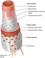
Most blood vessels (except capillaries) have three distinct layers, or tunics, that provide structural support and regulate vessel function:
Tunica intima: The innermost layer, composed of endothelium and elastic fibers, in direct contact with blood.
Tunica media: The middle layer, consisting of smooth muscle and elastic fibers, responsible for vasoconstriction and vasodilation.
Tunica externa (adventitia): The outermost layer, made of dense irregular connective tissue, prevents overstretching of the vessel.
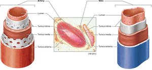
Comparison of Arteries and Veins
Arteries and veins differ in wall thickness, lumen size, and the presence of valves. Arteries have thicker walls and more elastic fibers to withstand higher pressure, while veins have larger lumens and valves to prevent backflow.
Types of Blood Vessels
Arteries
Arteries are classified into three main categories based on size and function:
Elastic arteries: Largest, thick-walled, closest to the heart (e.g., aorta); contain more elastic fibers and less smooth muscle.
Muscular arteries: Deliver blood to specific organs; have more smooth muscle for vasodilation/constriction (e.g., renal artery).
Arterioles: Smallest arteries with thin layers; regulate blood flow into capillaries.
Fun fact: Certain arteries contain baroreceptors and chemoreceptors to monitor blood pressure and blood chemistry.
Capillaries
Capillaries are the smallest and thinnest blood vessels, specialized for exchange between blood and tissues. Most tissues have a rich capillary supply to facilitate efficient exchange.
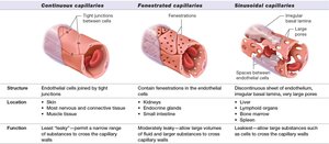
Types of Capillaries
Type | Structure | Location | Function |
|---|---|---|---|
Continuous | Endothelial cells joined by tight junctions | Skin, muscle, nervous tissue | Least leaky; restricts movement of substances |
Fenestrated | Endothelial cells with pores (fenestrations) | Kidneys, endocrine glands, small intestine | Moderately leaky; allows larger substances to cross |
Sinusoidal | Discontinuous endothelium, large pores | Liver, spleen, bone marrow | Most leaky; allows large substances and cells to cross |
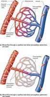
Capillary Beds and Microcirculation
Capillary beds are networks of capillaries interwoven through tissues. Microcirculation refers to blood flow through these beds, regulated by precapillary sphincters:
True capillaries: Open sphincters allow blood flow through the bed.
Vascular shunts: Closed sphincters direct blood through a shortcut channel.
Veins
Veins carry blood toward the heart, have all three tunics, and operate under low pressure. They contain valves to prevent backflow and have larger lumens than arteries. Capillaries merge to form venules, which then merge to form veins.
Fun fact: Veins are more numerous than arteries and contain about 70% of the body's blood volume.
Hemodynamics: Blood Flow and Pressure
Basic Principles
Hemodynamics is the study of blood flow and the forces involved. Blood flows from areas of higher pressure (near the heart) to areas of lower pressure (in the periphery). The pressure gradient is created by the heart's contraction.
Key Terms
Blood pressure (BP): The outward force exerted by blood on vessel walls; varies throughout the vasculature.
Blood flow (BF): The volume of blood moving through a vessel per minute; directly proportional to the pressure gradient.
Resistance (R): Any opposition to blood flow; inversely proportional to blood flow.

Peripheral Resistance
Peripheral resistance is the opposition to blood flow offered by the systemic blood vessels. It is influenced by vessel radius, blood viscosity, vessel length, and obstructions.
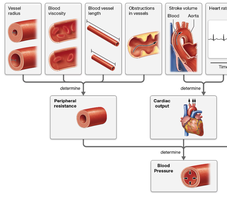
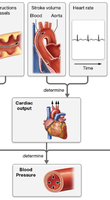
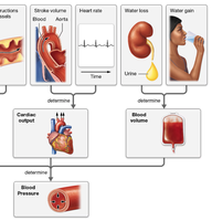
Cardiac Output and Blood Pressure
Cardiac output (CO) is the volume of blood pumped by the heart per minute, calculated as:
Stroke Volume (SV): Amount of blood pumped with each beat
Heart Rate (HR): Number of beats per minute
Formula:
The relationship between pressure, cardiac output, and resistance is:
Blood Volume and Compliance
Total blood volume is directly related to the amount of water in the blood. Small increases in blood volume are offset by vessel compliance (ability to stretch), especially in veins.
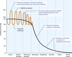
Blood Pressure in Different Areas
Arterial, Capillary, and Venous Pressure
Blood pressure is highest in the arteries and decreases as blood moves through arterioles, capillaries, venules, and veins. Systolic pressure occurs during ventricular contraction, while diastolic pressure occurs during relaxation.
Systolic pressure: 110–120 mm Hg
Diastolic pressure: 70–80 mm Hg
Mean Arterial Pressure (MAP): Average pressure in systemic arteries (~95 mm Hg)
Pulse Pressure: Difference between systolic and diastolic pressures (~40 mm Hg)

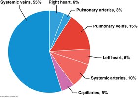
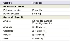
Circuit | Pressure |
|---|---|
Pulmonary arteries | 15 mm Hg |
Pulmonary veins | 5 mm Hg |
Systemic arteries | 120 mm Hg (systolic), 80 mm Hg (diastolic) |
Arterioles | 80–35 mm Hg |
Capillaries | 35–15 mm Hg |
Venules | 15–5 mm Hg |
Veins | 5–0 mm Hg |
Venous Return
Venous blood returns to the heart through mechanisms such as the skeletal muscle pump (in limbs) and the respiratory pump (in thoracic and abdominopelvic cavities). Veins contain valves to prevent backflow.
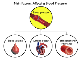
Regulation of Blood Pressure
Factors Affecting Blood Pressure
Blood pressure is determined by three main factors:
Cardiac output
Peripheral resistance
Blood volume
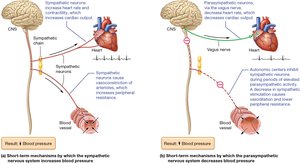
Short-Term Regulation: Nervous and Endocrine Systems
Short-term regulation involves rapid adjustments by the nervous and endocrine systems to maintain blood pressure through changes in heart rate, contractility, and vessel diameter.
Baroreceptor reflex: Baroreceptors in the carotid sinuses and aortic arch sense pressure changes and signal the medulla oblongata to adjust sympathetic or parasympathetic output.
Chemoreceptor stimulation: Chemoreceptors in the carotid and aortic bodies, and the medulla, sense changes in O2, CO2, and H+ and adjust cardiovascular activity accordingly.
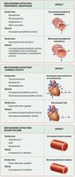
Key hormones involved in short-term regulation include:
Epinephrine/Norepinephrine (increase CO and PR)
Thyroid hormones (increase CO)
Angiotensin-II (increase PR)
Atrial Natriuretic Peptide (ANP) (decrease PR)
Long-Term Regulation: Renal Mechanisms
Long-term regulation of blood pressure is achieved by adjusting blood volume through the urinary system. The kidneys increase or decrease urine output to regulate blood volume and, consequently, blood pressure. Hormones such as angiotensin-II, ANP, antidiuretic hormone (ADH), and aldosterone play key roles in this process.
Disorders of Blood Vessels and Blood Pressure
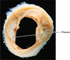
Atherosclerosis
Atherosclerosis is the formation of plaques (lipids, cholesterol, etc.) in the tunica intima of arteries, leading to narrowed vessels and increased risk of heart attack and stroke. Treatment includes lifestyle changes and, in severe cases, surgery.
Edema
Edema is the accumulation of excess fluid in the interstitial space, often caused by hypertension or liver failure.
Varicose Veins
Varicose veins are enlarged, twisted veins resulting from valve failure, commonly in the legs. They can cause discomfort and swelling.
Hypertension and Hypotension
Hypertension: Abnormally high blood pressure; risk factor for coronary artery disease, stroke, and heart failure. Most cases are idiopathic (unknown cause), but risk factors include genetics, age, diet, obesity, smoking, and alcohol use.
Hypotension: Abnormally low blood pressure; symptoms include dizziness, fainting, and organ dysfunction. Causes include low blood volume, decreased cardiac output, or excessive vasodilation.
Treatments for blood pressure disorders may include lifestyle changes, medications, or interventions to address underlying causes.
