 Back
BackThe Cardiovascular System: Blood Vessels and Circulation
Study Guide - Smart Notes
 Tailored notes based on your materials, expanded with key definitions, examples, and context.
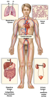
Cardiovascular System Overview
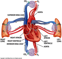
Pathway of Blood in the Body
The cardiovascular system is responsible for transporting blood throughout the body, delivering oxygen and nutrients to tissues, and removing waste products. Blood follows a specific pathway: heart → arteries → arterioles → capillaries → venules → veins → back to the heart.
Arteries carry blood away from the heart.
Veins return blood to the heart.
Capillaries are the sites of exchange between blood and tissues.
Structure of Blood Vessel Walls
General Organization
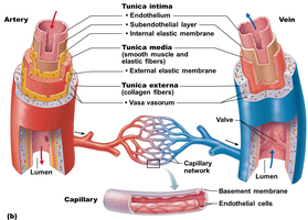
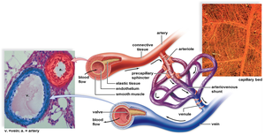
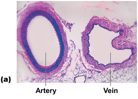
All blood vessels, except capillaries, have three layers (tunics) surrounding a central blood-containing space called the lumen:
Tunica intima: Innermost layer; consists of endothelium and a subendothelial layer.
Tunica media: Middle layer; composed of smooth muscle and elastic fibers, responsible for vasoconstriction and vasodilation.
Tunica externa: Outermost layer; made of collagen fibers that protect and anchor the vessel, contains the vasa vasorum (small vessels that supply the vessel wall).
Capillaries consist only of endothelium with a sparse basal lamina, allowing for efficient exchange of substances.
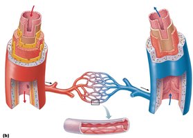
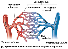
Vascular Anastomoses
Vascular anastomoses are interconnections between blood vessels, providing alternate pathways for blood flow in case of blockage.
Types of Blood Vessels
Arteries
Arteries carry blood away from the heart. Those closest to the heart (elastic arteries) have large diameters and thick walls to withstand and smooth out pressure fluctuations from the heart's pumping action. As arteries branch and move away from the heart, their diameter and wall thickness decrease.
Arterioles are small arteries that lead to capillary beds and regulate blood flow into tissues.
Capillaries
Capillaries are the smallest blood vessels, consisting of a single layer of endothelial cells. Their thin walls allow for rapid exchange of gases, nutrients, and wastes between blood and tissues.
Capillaries connect arterioles to venules.
They are present in almost all tissues except cartilage, epithelia, cornea, and lens of the eye.
Pericytes (spider-shaped stem cells) help stabilize capillary walls and control permeability.
Veins and Venules
Veins carry blood toward the heart. They form as capillary beds unite into postcapillary venules, which merge into larger veins. Veins have thinner walls and larger lumens than arteries, and often contain valves to prevent backflow, especially in the limbs.
Venules are small veins that collect blood from capillaries; they are very porous and allow fluids and white blood cells to pass into tissues.
Valves in veins prevent backflow of blood.
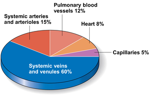
Distribution of Blood in the Body
Blood is unevenly distributed among the various vessels:
Location | Percentage of Blood Volume |
|---|---|
Systemic veins and venules | 60% |
Systemic arteries and arterioles | 15% |
Pulmonary blood vessels | 12% |
Heart | 8% |
Capillaries | 5% |
Physiology of Circulation
Key Terms and Concepts
Blood flow (F): Volume of blood flowing through a vessel, organ, or the entire circulation in a given period (ml/min). For the entire vascular system, it is equivalent to cardiac output (CO).
Blood pressure (BP): Force per unit area exerted on the wall of a blood vessel by the blood (measured in mm Hg).
Peripheral resistance (R): Opposition to blood flow due to friction between blood and vessel walls. Influenced by blood viscosity, vessel length, and vessel diameter.
The relationship between flow, pressure, and resistance is described by:
Where is blood flow, is the difference in blood pressure, and is resistance.
Systemic Blood Pressure
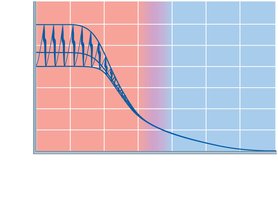
Blood pressure is highest in the aorta and decreases throughout the systemic circulation, with the steepest drop occurring in the arterioles.
Arterial Blood Pressure
Systolic pressure: Pressure in the aorta during ventricular contraction (e.g., 120 mm Hg).
Diastolic pressure: Lowest aortic pressure during heart relaxation (e.g., 80 mm Hg).
Pulse pressure: Difference between systolic and diastolic pressures.
Mean arterial pressure (MAP): Average pressure that propels blood to tissues. Calculated as:
Example: If BP = 120/80 mm Hg, Pulse Pressure = 40 mm Hg, so MAP = 80 + (1/3)×40 ≈ 93 mm Hg.
Venous Blood Pressure and Return
Factors Aiding Venous Return
Muscular pump: Skeletal muscle contractions "milk" blood toward the heart.
Respiratory pump: Pressure changes during breathing move blood toward the heart.
Sympathetic vasoconstriction: Smooth muscle constriction pushes blood toward the heart.
Regulation of Blood Pressure
Main Factors
Cardiac output (CO): Amount of blood pumped by the heart per minute.
Peripheral resistance (PR): Friction between blood and vessel walls.
Blood volume: Total amount of blood in the vascular system.
Blood pressure varies directly with CO, PR, and blood volume.
Short-Term Regulation: Neural Controls
Neural controls operate via reflex arcs involving baroreceptors, chemoreceptors, and higher brain centers.
Baroreceptor reflexes: Located in carotid sinuses, aortic arch, and large arteries; respond to changes in blood pressure by adjusting vessel diameter and heart rate.
Chemoreceptor reflexes: Respond to changes in blood CO2, pH, and O2 levels.
Higher brain centers (hypothalamus, cerebral cortex) can modify blood pressure during stress or exercise.
Short-Term Regulation: Hormonal Controls
Epinephrine and norepinephrine: Increase cardiac output and vasoconstriction.
Angiotensin II: Potent vasoconstrictor.
Antidiuretic hormone (ADH): High levels cause vasoconstriction and water retention.
Atrial natriuretic peptide (ANP): Decreases blood pressure by reducing blood volume.
Long-Term Regulation: Renal Mechanisms
Direct renal mechanism: Increased BP or blood volume leads to increased urine output, reducing BP; decreased BP causes kidneys to conserve water, raising BP.
Indirect renal mechanism (Renin-Angiotensin-Aldosterone System): Decreased BP triggers renin release, leading to formation of angiotensin II, which increases BP by vasoconstriction and stimulating aldosterone and ADH release.
Control of Blood Flow
Extrinsic and Intrinsic Regulation
Extrinsic control: Sympathetic nervous system and hormones regulate blood flow throughout the body, prioritizing vital organs.
Intrinsic (autoregulation) control: Local regulation of blood flow to meet tissue needs by adjusting arteriole diameter.
Developmental Aspects of Blood Vessels
Blood vessels develop from mesodermal cells forming blood islands, which become vascular tubes. Vascular endothelial growth factor determines vessel fate. Fetal shunts (foramen ovale, ductus arteriosus) bypass nonfunctional lungs. With aging, vascular problems such as varicose veins and atherosclerosis may occur.
