 Back
BackThe Cardiovascular System: Blood Vessels and Circulation
Study Guide - Smart Notes
 Tailored notes based on your materials, expanded with key definitions, examples, and context.
The Cardiovascular System: Blood Vessels and Circulation
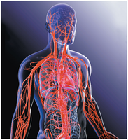
Introduction to Blood Vessels and Circuits
The cardiovascular system is composed of a complex network of blood vessels that transport blood throughout the body. There are two main circuits:
Pulmonary circuit: Carries blood to and from the lungs for gas exchange.
Systemic circuit: Delivers blood to the rest of the body and returns it to the heart.
Both circuits operate simultaneously with each heartbeat, ensuring efficient oxygen and nutrient delivery and waste removal.
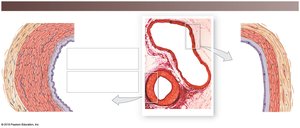
Histological Organization of Blood Vessels
General Structure of Vessel Walls
Except for capillaries, blood vessel walls consist of three layers (tunics):
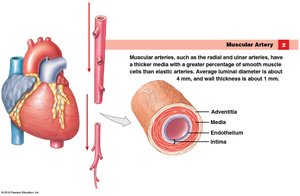
Adventitia (Tunica Externa): The outermost layer, primarily connective tissue, providing structural support and protection.
Media (Tunica Media): The middle layer, composed of smooth muscle and connective tissue, responsible for vasoconstriction and vasodilation.
Intima (Tunica Intima): The innermost layer, consisting of endothelium and connective tissue, providing a smooth lining for blood flow.
Comparison of Arteries and Veins
Arteries and veins can be distinguished by their wall structure and function:
Arteries: Carry blood away from the heart, have thicker walls, more smooth muscle, and greater elasticity to withstand higher pressure.
Veins: Carry blood toward the heart, have thinner walls, less smooth muscle, and larger lumens. Many veins contain valves to prevent backflow.
Types of Blood Vessels
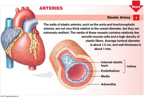
Arteries
As blood leaves the heart, it travels through arteries of decreasing diameter:
Elastic Arteries: Large vessels near the heart (e.g., aorta, brachiocephalic trunk). Highly elastic to accommodate pressure changes during the cardiac cycle.
Muscular Arteries: Medium-sized arteries (e.g., radial, ulnar, femoral). Thicker tunica media, regulate blood flow by vasoconstriction and vasodilation.
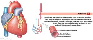
Arterioles: Smallest arteries, very thin walls, control blood flow into capillary beds.
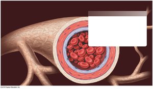
Capillaries
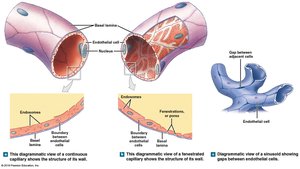
Capillaries are the smallest blood vessels and the primary site for exchange of nutrients, gases, and wastes between blood and tissues. They consist only of endothelium and a basal lamina.
Continuous Capillaries: No gaps between endothelial cells; found in most tissues.
Fenestrated Capillaries: Have pores (fenestrations) that allow greater permeability; found in kidneys, intestines, and endocrine glands.
Sinusoids: Large gaps and pores; found in liver, bone marrow, and spleen.
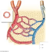
Capillary Beds
Capillaries form interconnected networks called capillary beds. Blood flow into capillary beds is regulated by precapillary sphincters. When these sphincters contract, blood bypasses the capillary bed via a thoroughfare channel.
Veins
Veins return blood to the heart and are classified by size:
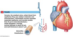
Venules: Smallest veins, collect blood from capillaries, thin or absent tunica media.
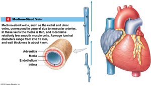
Medium-Sized Veins: Contain one-way valves, found alongside muscular arteries (e.g., radial, tibial veins).
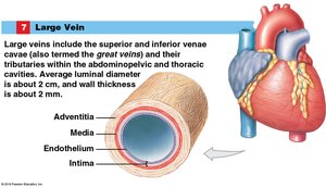
Large Veins: Include the superior and inferior vena cava, have thick walls but no valves.
The Pulmonary Circuit
Pathway of Blood Through the Pulmonary Circuit
The pulmonary circuit carries deoxygenated blood from the right ventricle to the lungs and returns oxygenated blood to the left atrium:
Right ventricle → pulmonary valve → pulmonary trunk
Pulmonary trunk → left and right pulmonary arteries
Pulmonary arteries → lungs (gas exchange: CO2 out, O2 in)
Pulmonary veins (4) → left atrium
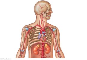
The Systemic Circuit
Major Systemic Arteries
Oxygenated blood leaves the left ventricle through the aortic valve into the ascending aorta, aortic arch, and descending aorta. Major branches include:
Brachiocephalic trunk: Right common carotid and right subclavian arteries
Left common carotid artery
Left subclavian artery
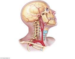
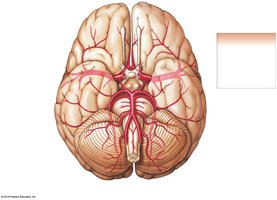
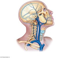
Circulation to the Brain
Blood supply to the brain is provided by the internal and external carotid arteries and vertebral arteries. The Cerebral Arterial Circle (Circle of Willis) ensures continuous blood flow to the brain.
Internal carotid → middle and anterior cerebral arteries
Vertebral arteries → basilar artery → posterior cerebral arteries
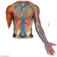
Arterial Supply to the Upper Limb
The subclavian artery continues as the axillary artery, then the brachial artery, which divides into the radial and ulnar arteries. These arteries anastomose at the wrist to form the superficial and deep palmar arches, supplying the hand and digits.
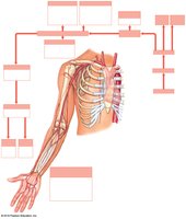
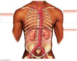
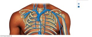
Arterial Supply to the Thorax and Abdomen
Branches of the subclavian and thoracic aorta supply the thorax, while the abdominal aorta gives rise to several paired and unpaired branches supplying abdominal organs.
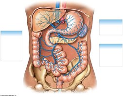
Unpaired branches: Celiac trunk, superior mesenteric, inferior mesenteric arteries
Paired branches: Inferior phrenic, suprarenal, renal, gonadal, lumbar arteries
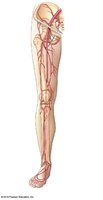
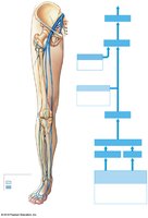
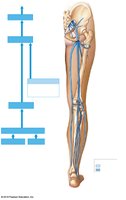
Arterial Supply to the Lower Limb
The abdominal aorta divides into the right and left common iliac arteries, which further branch to supply the pelvis and lower limbs (internal and external iliac, femoral, popliteal, tibial arteries).
Systemic Veins
Overview of Venous Return
Veins collect blood from tissues and return it to the heart via the superior and inferior vena cava. Venous drainage patterns can vary, and both deep and superficial veins are present in the limbs.

Venous Return from the Head, Neck, and Upper Limb
Blood from the head and neck drains into the internal and external jugular veins and vertebral veins, which empty into the brachiocephalic veins and then the superior vena cava. The upper limb is drained by superficial (cephalic, basilic) and deep (radial, ulnar, brachial) veins.
Venous Return from the Thorax and Abdomen
Intercostal veins drain into the azygos and hemiazygos veins, which empty into the superior vena cava. The inferior vena cava collects blood from the hepatic, phrenic, suprarenal, renal, gonadal, and lumbar veins. The hepatic portal system drains blood from the digestive organs to the liver before entering the inferior vena cava.
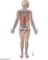
Venous Return from the Lower Limb
Blood from the lower limb is returned via the plantar and dorsal venous arches, which drain into the anterior and posterior tibial veins, popliteal vein, femoral vein, and ultimately the external and common iliac veins. The great and small saphenous veins are major superficial veins of the lower limb.
Summary Table: Comparison of Vessel Types
Vessel Type | Wall Structure | Function |
|---|---|---|
Elastic Artery | Thick tunica media, many elastic fibers | Conduct blood from heart, withstand pressure |
Muscular Artery | Thick tunica media, more smooth muscle | Distribute blood to organs, regulate flow |
Arteriole | Thin wall, little adventitia | Control blood flow to capillaries |
Capillary | Endothelium only | Exchange of gases, nutrients, wastes |
Venule | Thin wall, little or no media | Collect blood from capillaries |
Medium Vein | Thin media, valves present | Return blood to heart, prevent backflow |
Large Vein | Thick wall, no valves | Return blood to heart |
