 Back
BackThe Cardiovascular System: Blood Vessels – Structure, Function, and Regulation
Study Guide - Smart Notes
 Tailored notes based on your materials, expanded with key definitions, examples, and context.
Blood Vessel Structure and Function
Overview of the Vascular System
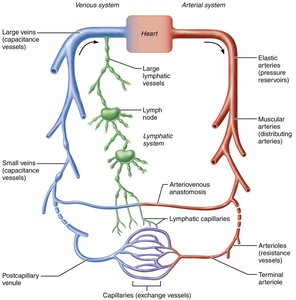
The cardiovascular system is composed of a closed network of blood vessels that transport blood throughout the body, working in conjunction with the lymphatic system to maintain fluid balance and tissue health. Blood vessels are dynamic structures that can constrict, dilate, and adapt to physiological demands.
Arteries: Carry blood away from the heart. Systemic arteries transport oxygenated blood, while pulmonary arteries carry oxygen-poor blood.
Capillaries: Serve as exchange vessels, allowing for the transfer of gases, nutrients, and wastes between blood and tissues.
Veins: Return blood to the heart. Systemic veins carry deoxygenated blood, while pulmonary veins carry oxygenated blood.
Blood Vessel Anatomy
Layers of Blood Vessel Walls
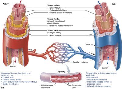
Except for capillaries, most blood vessels have three distinct layers (tunics) surrounding a central lumen:
Tunica intima: Innermost layer, composed of endothelium (simple squamous epithelium) and a subendothelial layer in larger vessels. Provides a smooth, friction-reducing lining.
Tunica media: Middle layer, primarily smooth muscle and elastic fibers. Responsible for vasoconstriction and vasodilation, thus regulating blood flow and pressure.
Tunica externa (adventitia): Outermost layer, mainly collagen fibers that protect, reinforce, and anchor the vessel. Contains nerves, lymphatics, and in large vessels, vasa vasorum (small vessels that nourish the outer wall).
Capillaries consist only of endothelium and a sparse basal lamina, facilitating efficient exchange.
Comparative Anatomy of Blood Vessels
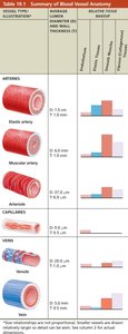
Arteries, veins, and capillaries differ in structure and function. The following table summarizes their key anatomical features:
Vessel Type | Diameter | Wall Thickness | Key Features |
|---|---|---|---|
Elastic artery | 1.0–2.5 cm | 1.0–1.5 mm | Thick tunica media, abundant elastin, pressure reservoir |
Muscular artery | 0.3 mm–1.0 cm | 0.5–1.0 mm | Thickest tunica media, more smooth muscle, distributing vessel |
Arteriole | 10–300 μm | 6–30 μm | Smallest arteries, control flow into capillaries, resistance vessels |
Capillary | 8–10 μm | 0.5 μm | Single endothelial layer, exchange vessel |
Venule | 8–100 μm | 1–2 μm | Very porous, allow fluid and WBC movement |
Vein | 0.1 mm–2.5 cm | 0.5 mm | Thin walls, large lumen, valves present, capacitance vessel |
Histological Comparison: Arteries vs. Veins
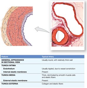
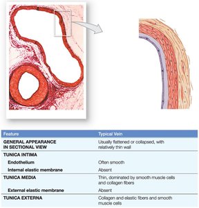
Arteries and veins can be distinguished by their wall structure and appearance in cross-section:
Arteries: Thick walls, round lumen, prominent tunica media with smooth muscle and elastic fibers.
Veins: Thinner walls, often collapsed or irregular lumen, less smooth muscle, valves may be present.
Types of Capillaries
Continuous Capillaries
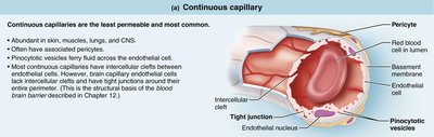
Continuous capillaries are the most common and least permeable type, found in skin, muscles, lungs, and the central nervous system. They have tight junctions between endothelial cells but allow limited passage of fluids and small solutes through intercellular clefts.
Fenestrated Capillaries
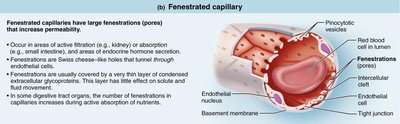
Fenestrated capillaries contain pores (fenestrations) that increase permeability. They are found in areas of active filtration (kidneys), absorption (intestines), and endocrine hormone secretion.
Sinusoidal Capillaries
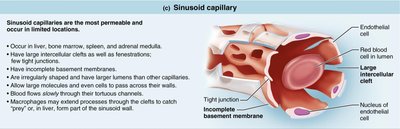
Sinusoidal capillaries are the most permeable and are found in the liver, bone marrow, spleen, and adrenal medulla. They have large intercellular clefts, fenestrations, and an incomplete basement membrane, allowing the passage of large molecules and cells.
Capillary Beds and Microcirculation
Capillary Bed Structure and Regulation
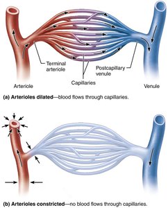
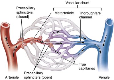
Capillary beds are networks of capillaries between arterioles and venules, facilitating exchange with tissues. Blood flow through these beds is regulated by the diameter of arterioles and the action of precapillary sphincters, which respond to local chemical conditions.
Veins: Structure and Function
Venous System and Blood Reservoirs
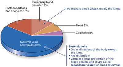
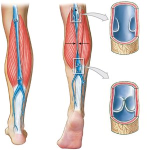
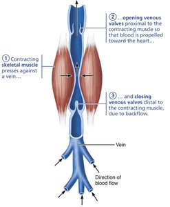
Veins return blood to the heart and serve as blood reservoirs, containing up to 65% of the blood volume at any time. They have thinner walls and larger lumens than arteries, and their low pressure requires adaptations to ensure blood return to the heart, such as valves and the muscular pump.
Blood Flow, Pressure, and Resistance
Key Hemodynamic Principles
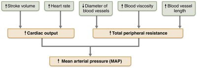
Blood flow is the volume of blood moving through a vessel, organ, or the entire circulation per unit time. Blood pressure is the force per unit area exerted on a vessel wall by the blood, and resistance is the opposition to flow, primarily due to friction within the vessel.
Blood flow (F) is directly proportional to the pressure gradient (ΔP) and inversely proportional to total peripheral resistance (TPR):
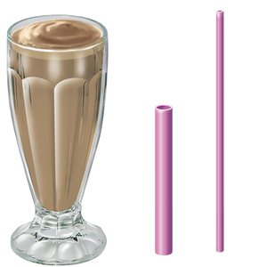
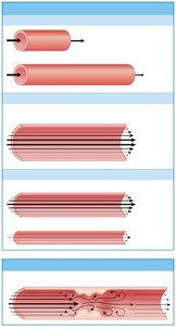
Resistance is affected by blood viscosity, vessel length, and especially vessel diameter (inversely proportional to the fourth power of the radius).
Blood Pressure Throughout the Circulation
Pressure Changes in the Systemic Circuit
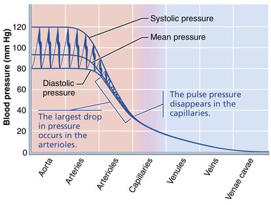
Blood pressure is highest in the aorta and declines through the systemic circuit, with the steepest drop in the arterioles. Capillary pressure is low to prevent rupture and allow exchange, while venous pressure is steady and low.
Measuring Blood Pressure and Pulse
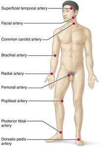
Blood pressure is measured using a sphygmomanometer, typically at the brachial artery. Systolic pressure is the peak during ventricular contraction, and diastolic is the lowest during relaxation. Pulse pressure is the difference between systolic and diastolic pressures. Mean arterial pressure (MAP) is calculated as:
Regulation of Blood Pressure
Short- and Long-Term Controls
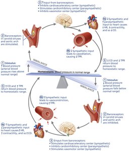
Blood pressure is regulated by cardiac output (CO), total peripheral resistance (TPR), and blood volume. Short-term controls involve neural and hormonal mechanisms that alter vessel diameter and heart function, while long-term controls involve renal mechanisms that adjust blood volume.
Neural controls: Baroreceptor and chemoreceptor reflexes, cardiovascular center in the medulla.
Hormonal controls: Epinephrine, norepinephrine, angiotensin II, ADH, aldosterone, and ANP.
Renal controls: Direct (filtration) and indirect (renin-angiotensin-aldosterone system) mechanisms.
Capillary Exchange and Bulk Flow
Mechanisms of Exchange
Capillaries allow the exchange of gases, nutrients, and wastes via diffusion, vesicular transport, and bulk flow. Bulk flow is driven by hydrostatic and osmotic pressures, determining the direction and amount of fluid movement.
Hydrostatic pressure: Pushes fluid out of capillaries (filtration).
Colloid osmotic pressure: Pulls fluid into capillaries (reabsorption).
Net filtration pressure (NFP):
Clinical Correlations
Homeostatic Imbalances
Hypertension: Chronic high blood pressure, risk factor for heart disease, stroke, and kidney failure.
Hypotension: Abnormally low blood pressure, may cause inadequate tissue perfusion.
Edema: Excess interstitial fluid due to increased filtration or decreased reabsorption, can result from heart failure, inflammation, or lymphatic obstruction.
Circulatory Pathways
Pulmonary and Systemic Circulation
The vascular system consists of pulmonary circulation (heart to lungs and back) and systemic circulation (heart to body tissues and back). Systemic arteries and veins differ in their pathways and anatomical relationships.
Summary Table: Blood Vessel Types and Functions
Vessel Type | Main Function | Key Structural Feature |
|---|---|---|
Elastic artery | Pressure reservoir | Thick tunica media, abundant elastin |
Muscular artery | Distributing vessel | Thick smooth muscle, less elastic tissue |
Arteriole | Resistance vessel | Small diameter, smooth muscle |
Capillary | Exchange vessel | Single endothelial layer |
Venule | Collecting vessel | Porous, thin wall |
Vein | Capacitance vessel | Large lumen, valves |
