 Back
BackThe Cardiovascular System: Blood Vessels – Structure and Function
Study Guide - Smart Notes
 Tailored notes based on your materials, expanded with key definitions, examples, and context.
The Cardiovascular System: Blood Vessels
Introduction to Blood Vessels
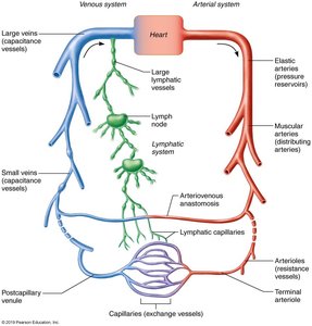
The cardiovascular system is composed of a dynamic network of blood vessels that transport blood throughout the body. Blood vessels work closely with the lymphatic system to maintain fluid balance and deliver essential substances to tissues.
Arteries: Carry blood away from the heart; usually oxygenated except in pulmonary circulation and fetal vessels.
Capillaries: Microscopic vessels that directly serve tissue cells, facilitating exchange of gases, nutrients, and wastes.
Veins: Return blood to the heart; usually deoxygenated except in pulmonary circulation and fetal vessels.
Structure of Blood Vessel Walls
General Organization
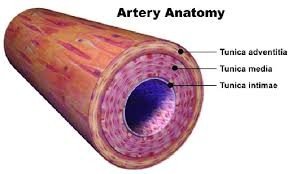
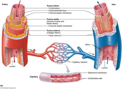
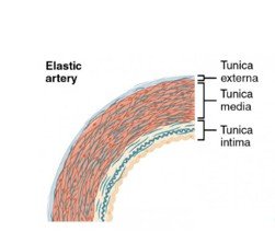
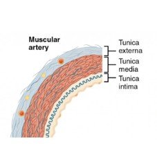
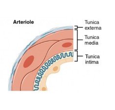
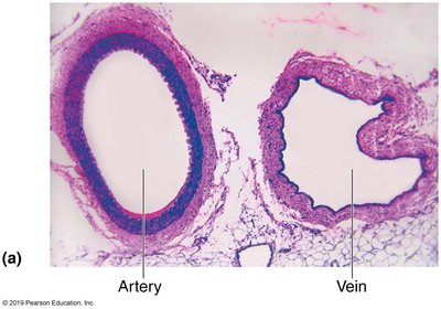
All blood vessels (except capillaries) share a similar wall structure composed of three layers, or tunics, surrounding a central lumen:
Tunica intima: Innermost layer, consisting of endothelium (simple squamous epithelium) and, in larger vessels, a subendothelial connective tissue layer.
Tunica media: Middle layer, primarily smooth muscle and elastin, responsible for vasoconstriction and vasodilation.
Tunica externa (adventitia): Outermost layer, mainly loose collagen fibers that protect, reinforce, and anchor the vessel. Contains nerves, lymphatics, and in large vessels, the vasa vasorum (small blood vessels that nourish the outer wall).
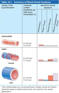
Summary Table: Blood Vessel Anatomy
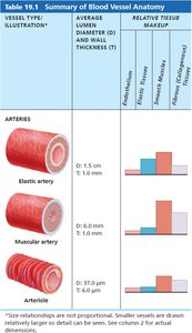
The following table summarizes the main structural differences among arteries, capillaries, and veins:
Vessel Type | Diameter & Wall Thickness | Key Features |
|---|---|---|
Elastic artery | D: 1.5 cm, T: 1.0 mm | High elastic fibers, thick wall, pressure reservoir |
Muscular artery | D: 6.0 mm, T: 1.0 mm | Thickest smooth muscle, distributing arteries |
Arteriole | D: 37.0 μm, T: 6.0 μm | Smallest arteries, regulate blood flow into capillaries |
Capillary | D: 9.0 μm, T: 0.5 μm | Single endothelial layer, exchange vessels |
Venule | D: 20.0 μm, T: 1.0 μm | Very porous, allow fluid and WBC movement |
Vein | D: 5.0 mm, T: 0.5 mm | Thin walls, large lumen, valves present |
Types of Arteries
Elastic Arteries
Elastic arteries are the largest arteries, including the aorta and its major branches. They have thick walls with abundant elastic fibers, allowing them to stretch and recoil as blood is ejected from the heart. This property enables them to act as pressure reservoirs, maintaining continuous blood flow even between heartbeats.
Inactive in vasoconstriction; mainly function to conduct blood.
Muscular Arteries
Muscular arteries, also known as distributing arteries, deliver blood to specific body organs. They have the thickest tunica media with more smooth muscle and less elastic tissue, making them active in vasoconstriction and regulation of blood flow.
Arterioles
Arterioles are the smallest arteries, leading directly to capillary beds. They have a thin muscle layer and are the primary regulators of blood pressure before blood enters the capillaries. Their diameter changes in response to neural, hormonal, and local chemical signals, controlling blood flow into tissues.
Capillaries
Structure and Function
Capillaries are microscopic vessels with walls consisting of a single layer of endothelial cells and a sparse basal lamina. Their small diameter allows only one red blood cell to pass at a time. Capillaries are the primary sites for exchange of gases, nutrients, wastes, and hormones between blood and interstitial fluid.
Pericytes: Support cells that stabilize capillary walls and regulate permeability.
Types of Capillaries
Continuous capillaries: Most common; found in skin, muscles, lungs, and CNS. Endothelial cells are joined by tight junctions with small intercellular clefts for limited permeability. In the brain, they form the blood-brain barrier.
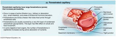
Fenestrated capillaries: Have pores (fenestrations) that increase permeability; found in kidneys, intestines, and endocrine glands.
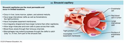
Sinusoidal capillaries: Most permeable, with large gaps and incomplete basement membranes; found in liver, bone marrow, spleen, and adrenal medulla. Allow passage of large molecules and cells.
Capillary Beds and Microcirculation
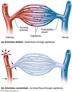
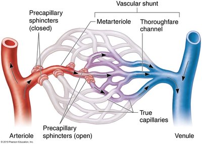
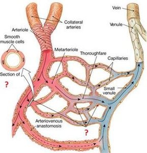
Capillary beds are networks of capillaries between arterioles and venules. Blood flow through these beds is regulated by the diameter of arterioles and local chemical conditions. Precapillary sphincters control the entry of blood into true capillaries, allowing for regulation of tissue perfusion.
Veins and Venules
Structure and Function
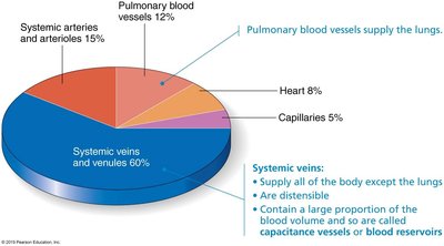
Veins carry blood toward the heart and are formed by the convergence of venules. They have thinner walls and larger lumens compared to arteries, making them effective blood reservoirs (capacitance vessels). Veins contain up to 65% of the body's blood supply.
Blood pressure in veins is lower than in arteries.
Adaptations for venous return include large lumens and valves that prevent backflow, especially in the limbs.
Venous sinuses are specialized, flattened veins with very thin walls (e.g., coronary sinus, dural sinuses).
Clinical Application: Varicose Veins and Hemorrhoids
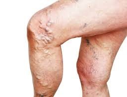
Varicose veins are dilated, twisted veins resulting from incompetent (leaky) valves. Factors such as heredity, prolonged standing, obesity, and pregnancy can contribute to their development. Elevated venous pressure, such as from straining, can also cause varicosities in anal veins, known as hemorrhoids.
Vascular Anastomoses
Types and Functions
Vascular anastomoses are interconnections between blood vessels that provide alternate pathways for blood flow. They are crucial for ensuring continuous circulation even if one pathway is blocked.
Arterial anastomoses: Common in joints, abdominal organs, brain, and heart; absent in retina, kidneys, and spleen.
Arteriovenous anastomoses: Direct connections between arterioles and venules, bypassing capillaries; important for thermoregulation in skin and extremities.
Venous anastomoses: Very abundant, ensuring that blocked veins rarely impede blood return.
Summary Table: Blood Vessel Structure and Function
Vessel Type | Main Features | Function |
|---|---|---|
Elastic arteries | Thick walls, high elastic fiber content | Pressure reservoirs, conduct blood from heart |
Muscular arteries | Thick smooth muscle, less elastic tissue | Distribute blood to organs, regulate flow |
Arterioles | Smallest arteries, thin muscle layer | Regulate blood flow into capillaries |
Capillaries | Single endothelial layer | Exchange of gases, nutrients, wastes |
Venules | Porous, few muscle cells | Collect blood from capillaries |
Veins | Thin walls, large lumens, valves | Return blood to heart, blood reservoirs |
Key Terms and Concepts
Vasoconstriction: Decrease in vessel diameter, increasing resistance and blood pressure.
Vasodilation: Increase in vessel diameter, decreasing resistance and blood pressure.
Vasa vasorum: Small vessels that supply blood to the walls of large arteries and veins.
Precapillary sphincter: Smooth muscle ring regulating blood flow into capillaries.
Blood-brain barrier: Specialized continuous capillaries in the CNS with tight junctions and no clefts.
Additional info: This guide covers the structure and function of blood vessels, including clinical relevance and anatomical variations, as outlined in Chapter 19 of a standard Human Anatomy & Physiology textbook.
