 Back
The Cardiovascular System: Blood Vessels – Structure, Function, and Physiology
Study Guide - Smart Notes
 Tailored notes based on your materials, expanded with key definitions, examples, and context.
Blood Vessel Structure and Function
Overview of the Vascular System
The cardiovascular system is a closed delivery system of blood vessels that begins and ends at the heart. Blood vessels are dynamic structures that pulsate, constrict, relax, and even multiply, working closely with the lymphatic system to circulate fluids throughout the body.
Arteries: Carry blood away from the heart toward capillaries. Systemic arteries transport oxygenated blood, while pulmonary arteries carry oxygen-poor blood. Umbilical arteries transport oxygen-poor blood from the fetus to the placenta.
Capillaries: Serve as exchange vessels, allowing substances to move between blood and tissue cells.
Veins: Carry blood from capillaries back toward the heart. Systemic veins carry oxygen-poor blood; pulmonary veins carry oxygenated blood. The umbilical vein transports oxygenated blood from the placenta to the fetus.
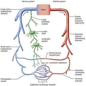
Additional info: The lymphatic system collects excess tissue fluid and returns it to the bloodstream, maintaining fluid balance.
Blood Vessel Wall Structure
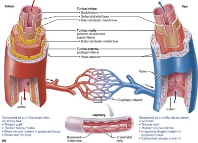
Most blood vessel walls (except capillaries) have three layers, or tunics, surrounding a central lumen:
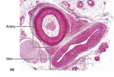
Tunica intima: Innermost layer, composed of endothelium (simple squamous epithelium) and a subendothelial layer in larger vessels. Provides a smooth, friction-reducing lining.
Tunica media: Middle layer, mostly smooth muscle and elastin. Regulated by sympathetic vasomotor nerve fibers, allowing for vasoconstriction (decreased lumen diameter) and vasodilation (increased lumen diameter). Responsible for maintaining blood flow and pressure.
Tunica externa (adventitia): Outermost layer, mainly collagen fibers that protect, reinforce, and anchor the vessel. Contains nerve fibers, lymphatic vessels, and in large vessels, the vasa vasorum ("vessels of the vessels") that nourish the outer wall.
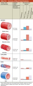
Summary Table: Blood Vessel Anatomy
The following table summarizes the main structural and functional differences among the major types of blood vessels:
Type | Diameter (mm) | Wall Thickness (mm) | Tunica Media | Relative Elasticity | Relative Muscle Content |
|---|---|---|---|---|---|
Elastic artery | 1.0–2.5 | 1.0 | Thick | High | Moderate |
Muscular artery | 0.3–1.0 | 1.0 | Thicker | Moderate | High |
Arteriole | 0.01–0.3 | 0.6 | Thin | Low | Moderate |
Capillary | 0.008–0.01 | 0.5 | None | None | None |
Venule | 0.02–0.5 | 0.5 | Very thin | None | Low |
Vein | 0.5–5.0 | 0.5 | Thin | Low | Low |
Types of Blood Vessels
Arteries
Elastic arteries: Thick-walled, near the heart (e.g., aorta). Act as pressure reservoirs, expanding and recoiling to maintain blood flow during the cardiac cycle. Diseases like atherosclerosis can stiffen these arteries, leading to aneurysms.
Muscular arteries: Distribute blood to specific organs. Have the thickest tunica media, more smooth muscle, and less elastic tissue than elastic arteries. More active in vasoconstriction.
Arterioles: Smallest arteries, leading into capillary beds. Control blood flow into capillaries via vasodilation and vasoconstriction. Major determinants of total peripheral resistance (TPR).
Capillaries
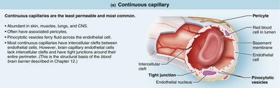
Capillaries are the smallest blood vessels, consisting only of a thin tunica intima. Their primary function is the exchange of gases, nutrients, wastes, and hormones between blood and interstitial fluid. Most tissues have a rich capillary supply, except for poorly vascularized tendons, ligaments, cartilage, and epithelia.
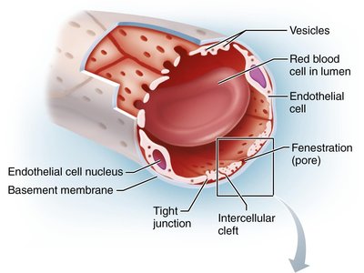
Continuous capillaries: Least permeable and most common. Found in skin, muscles, lungs, and CNS. Endothelial cells joined by tight junctions with intercellular clefts for limited passage of fluids and small solutes.
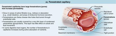
Fenestrated capillaries: Have pores (fenestrations) that increase permeability. Found in kidneys, intestines, and endocrine glands—sites of active filtration or absorption.
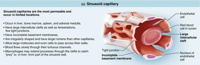
Sinusoidal capillaries: Most permeable, with large intercellular clefts and fenestrations. Found in liver, bone marrow, spleen, and adrenal medulla. Allow passage of large molecules and cells.
Capillary Beds
Capillary beds are interwoven networks of capillaries between arterioles and venules, providing sites for exchange between blood and tissues. Blood flow through capillary beds is regulated by the diameter of arterioles and local chemical conditions.
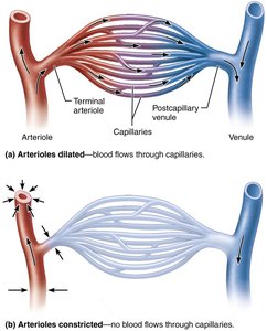
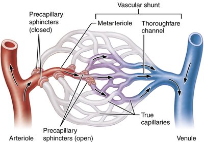
In some tissues, such as the intestinal mesenteries, capillary beds have additional features:
Vascular shunt: Directly connects terminal arteriole to postcapillary venule, bypassing true capillaries.
Precapillary sphincters: Rings of smooth muscle that regulate blood flow into true capillaries, controlled by local chemical conditions.
Veins and Venules
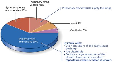
Veins carry blood toward the heart and act as blood reservoirs, containing up to 65% of the blood supply at any time. Venules are formed by the union of capillaries and are very porous, allowing fluids and white blood cells to move into tissues. Veins have thinner walls and larger lumens than arteries, with adaptations such as valves to prevent backflow and venous sinuses for specialized drainage.
Physiology of Circulation
Blood Flow, Pressure, and Resistance
Blood flow (F): Volume of blood flowing through a vessel, organ, or the entire circulation per minute (ml/min). Equivalent to cardiac output (CO) for the entire vascular system.
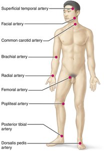
Blood pressure (BP): Force per unit area exerted on a vessel wall by the blood, measured in mm Hg. Systemic arterial BP is measured in large arteries near the heart.
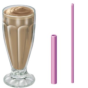
Resistance (R or TPR): Opposition to flow, mainly due to friction between blood and vessel walls. Determined by blood viscosity, vessel length, and vessel diameter.
Key relationships:
Blood flow is directly proportional to the pressure gradient () and inversely proportional to resistance ():
Vessel diameter is the most important factor affecting resistance, as resistance varies inversely with the fourth power of the radius:
Blood Pressure Changes Throughout the Circulation
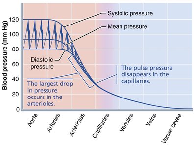
Blood pressure is highest in the aorta and declines throughout the systemic circulation, reaching 0 mm Hg in the right atrium. The steepest drop occurs in the arterioles, where resistance is greatest.
Arterial Blood Pressure
Systolic pressure: Pressure during ventricular contraction (average 120 mm Hg).
Diastolic pressure: Lowest pressure during ventricular relaxation (average 70–80 mm Hg).
Pulse pressure: Difference between systolic and diastolic pressure.
Mean arterial pressure (MAP): Average pressure that propels blood to tissues. Calculated as:
Pulse and MAP decline with distance from the heart.
Venous Blood Pressure and Return
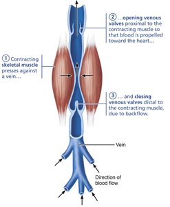
Venous pressure is steady and low, requiring adaptations to return blood to the heart:
Muscular pump: Skeletal muscle contractions "milk" blood toward the heart.
Respiratory pump: Pressure changes during breathing move blood toward the heart.
Sympathetic venoconstriction: Reduces vein capacity, pushing blood toward the heart.
Regulation of Blood Pressure
Major Factors Affecting Blood Pressure
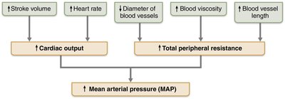
Cardiac output (CO)
Total peripheral resistance (TPR)
Blood volume
Blood pressure varies directly with CO, TPR, and blood volume:
Since , then:
Short-Term Regulation
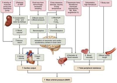
Neural controls: Baroreceptor reflexes (monitor stretch), chemoreceptor reflexes (monitor CO2, H+, O2), and higher brain centers regulate vessel diameter and blood distribution.
Hormonal controls: Epinephrine, norepinephrine, angiotensin II, ADH, aldosterone, and ANP affect blood pressure by altering CO, TPR, or blood volume.
Long-Term Regulation: Renal Mechanisms
Direct mechanism: Kidneys adjust blood volume by eliminating or conserving water.
Indirect mechanism: Renin-angiotensin-aldosterone system increases blood volume and vasoconstriction in response to low blood pressure.
Blood Flow Through Tissues
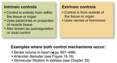
Intrinsic vs. Extrinsic Controls
Intrinsic (autoregulation): Local regulation of blood flow to match tissue metabolic needs.
Extrinsic: Neural and hormonal mechanisms maintain systemic blood pressure and redistribute blood as needed.
Special Circulations
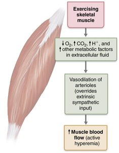
Skeletal muscle: Blood flow increases with activity (active hyperemia).
Capillary Exchange and Bulk Flow
Velocity of Blood Flow
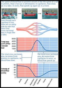
Blood flow is fastest in the aorta, slowest in capillaries (allowing exchange), and increases again in veins. Speed is inversely related to total cross-sectional area.
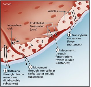
Capillary Exchange Mechanisms
Substances move between blood and interstitial fluid by:
Diffusion through endothelial membranes (lipid-soluble substances)
Movement through intercellular clefts or fenestrations (water-soluble substances)
Vesicular transport (large molecules)
Bulk Flow: Hydrostatic and Osmotic Pressures
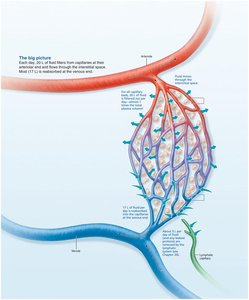
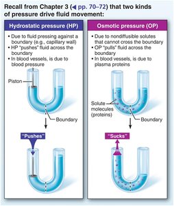
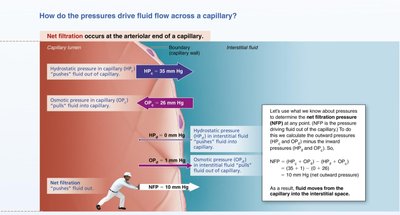
Bulk flow of fluid across capillary walls is determined by hydrostatic pressure (pushes fluid out) and colloid osmotic pressure (pulls fluid in). Net filtration pressure (NFP) determines the direction of fluid movement:
At the arterial end, NFP is positive (filtration).
At the venous end, NFP is negative (reabsorption).
Excess fluid is returned to the blood via the lymphatic system.
Clinical Correlations
Homeostatic Imbalances
Hypertension: Chronic high blood pressure, a major risk factor for heart failure, stroke, and kidney disease.
Hypotension: Low blood pressure, usually not a concern unless it causes inadequate tissue perfusion.
Circulatory shock: Any condition where blood vessels are inadequately filled, leading to poor tissue perfusion (e.g., hypovolemic, vascular, or cardiogenic shock).
Edema: Abnormal increase in interstitial fluid, caused by increased capillary hydrostatic pressure, decreased plasma proteins, or lymphatic obstruction.
Summary
The cardiovascular system's blood vessels are specialized for transport, exchange, and regulation of blood flow and pressure. Understanding their structure and function is essential for comprehending how the body maintains homeostasis and responds to physiological and pathological changes.
