 Back
The Cardiovascular System: Blood Vessels – Structure, Function, and Physiology
Study Guide - Smart Notes
 Tailored notes based on your materials, expanded with key definitions, examples, and context.
Blood Vessel Structure and Function
Overview of Blood Vessels
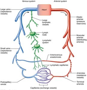
The cardiovascular system is composed of a dynamic network of blood vessels that transport blood throughout the body. These vessels work closely with the lymphatic system to maintain fluid balance and support cellular needs.
Arteries: Carry blood away from the heart; typically oxygenated except in pulmonary circulation and fetal umbilical vessels.
Capillaries: Microscopic vessels that directly serve tissue cells, facilitating exchange of gases, nutrients, and wastes.
Veins: Return blood toward the heart; typically deoxygenated except in pulmonary circulation and fetal umbilical vessels.
General Structure of Blood Vessel Walls
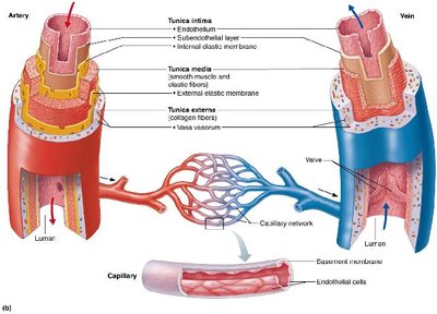
Except for capillaries, all blood vessels have three distinct layers (tunics) surrounding a central lumen:
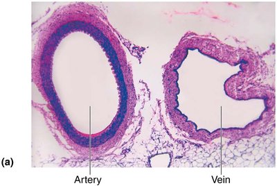
Tunica intima: Innermost layer, composed of endothelium (simple squamous epithelium) and, in larger vessels, a subendothelial connective tissue layer. Provides a smooth surface to reduce friction.
Tunica media: Middle layer, primarily smooth muscle and elastin. Responsible for vasoconstriction (narrowing) and vasodilation (widening) of the vessel, thus regulating blood flow and pressure.
Tunica externa (adventitia): Outermost layer, mainly loose collagen fibers that protect, reinforce, and anchor the vessel. Contains nerves, lymphatics, and in large vessels, the vasa vasorum (small blood vessels that nourish the outer wall).
Summary Table: Blood Vessel Anatomy
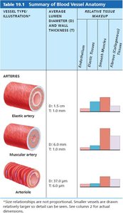
The following tables summarize the structural differences among the major vessel types:
Vessel Type | Diameter (D) | Wall Thickness (T) | Endothelium | Elastic Fibers | Smooth Muscle | Collagen Fibers |
|---|---|---|---|---|---|---|
Elastic artery | 1.5 cm | 1.0 mm | ++ | +++ | ++ | + |
Muscular artery | 6.0 mm | 1.0 mm | ++ | + | +++ | + |
Arteriole | 37.0 μm | 6.0 μm | + | - | + | - |
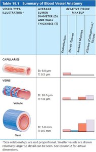
Vessel Type | Diameter (D) | Wall Thickness (T) | Endothelium | Elastic Fibers | Smooth Muscle | Collagen Fibers |
|---|---|---|---|---|---|---|
Capillary | 9.0 μm | 0.5 μm | + | - | - | - |
Venule | 20.0 μm | 1.0 μm | + | - | - | + |
Vein | 5.0 mm | 0.5 mm | + | + | + | ++ |
Types of Arteries
Elastic (Conducting) Arteries
These are the largest arteries (e.g., aorta and its major branches) and act as pressure reservoirs, expanding and recoiling to maintain continuous blood flow during the cardiac cycle. They contain abundant elastin in all three tunics but are relatively inactive in vasoconstriction.
Function: Conduct blood from the heart to medium-sized arteries.
Muscular (Distributing) Arteries
These arteries deliver blood to specific organs and have the thickest tunica media, with more smooth muscle and less elastic tissue than elastic arteries. They are active in vasoconstriction and vasodilation.
Function: Distribute blood to body organs and regulate blood flow by changing vessel diameter.
Arterioles
The smallest arteries, arterioles control blood flow into capillary beds through vasoconstriction and vasodilation. They are the primary resistance vessels, as their diameter changes significantly affect blood flow and resistance.
Capillaries: Structure and Types
General Structure
Capillaries are composed of a single layer of endothelial cells and a sparse basal lamina. Their thin walls facilitate the exchange of substances between blood and interstitial fluid.
Types of Capillaries
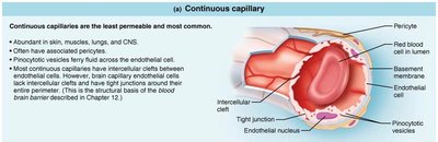
Continuous Capillaries: Least permeable and most common; found in skin, muscles, lungs, and CNS. Endothelial cells are joined by tight junctions with intercellular clefts for limited passage of fluids and small solutes. In the brain, they form the blood-brain barrier.
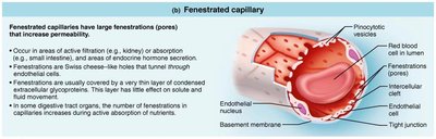
Fenestrated Capillaries: Have pores (fenestrations) that increase permeability; found in areas of active filtration (kidneys), absorption (intestines), or endocrine hormone secretion.
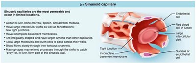
Sinusoidal Capillaries: Most permeable, with large intercellular clefts and incomplete basement membranes; found in the liver, bone marrow, spleen, and adrenal medulla. Allow passage of large molecules and cells; contain macrophages for immune defense.
Capillary Beds and Microcirculation
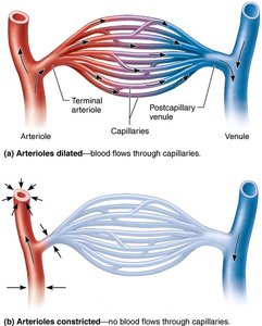
Capillary beds are networks of capillaries between arterioles and venules. Blood flow through these beds is regulated by the diameter of arterioles and local chemical conditions.
Precapillary sphincters control blood flow into true capillaries.
Vascular shunt (metarteriole-thoroughfare channel) allows blood to bypass true capillaries when sphincters are closed.
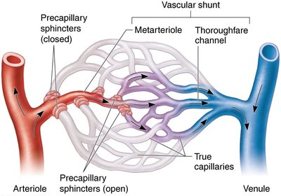
Veins: Structure and Function
General Structure
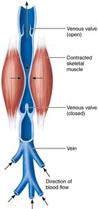
Veins have the same three tunics as arteries but with thinner walls and larger lumens. The tunica externa is the thickest layer, and veins often contain valves to prevent backflow, especially in the limbs.
Venules are formed when capillaries unite; they are very porous and allow fluids and white blood cells to pass into tissues.
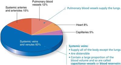
Veins act as blood reservoirs, containing up to 65% of the body's blood volume.
Blood Volume Distribution
Most blood is found in the systemic veins and venules, which serve as capacitance vessels or blood reservoirs.
Physiology of Circulation
Blood Flow, Pressure, and Resistance
Blood flow is the volume of blood moving through a vessel, organ, or the entire circulation per unit time (ml/min). Blood pressure is the force per unit area exerted by blood on vessel walls, and resistance is the opposition to flow, mainly due to friction within the vessel.
Key equation: , where is blood flow, is the pressure gradient, and is resistance.
Resistance is most affected by vessel diameter (inversely proportional to the fourth power of the radius).
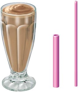
Systemic Blood Pressure
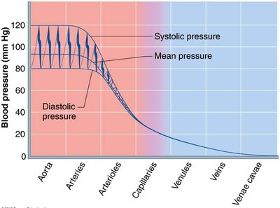
Blood pressure is highest in the aorta and decreases through the systemic circuit, with the steepest drop in arterioles. Systolic pressure is the peak during ventricular contraction, and diastolic is the lowest during relaxation. Mean arterial pressure (MAP) is the average pressure driving blood to tissues.
MAP calculation:
Venous Return Mechanisms
Because venous pressure is low, several mechanisms assist blood return to the heart:
Muscular pump: Skeletal muscle contractions squeeze veins, pushing blood toward the heart.
Respiratory pump: Pressure changes during breathing move blood toward the heart.
Venoconstriction: Sympathetic stimulation reduces vein diameter, increasing venous return.
Regulation of Blood Pressure
Major Factors Affecting MAP
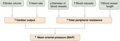
Mean arterial pressure (MAP) is determined by cardiac output (CO), total peripheral resistance (TPR), and blood volume. Changes in stroke volume, heart rate, vessel diameter, blood viscosity, and vessel length all influence MAP.
Neural and Hormonal Controls
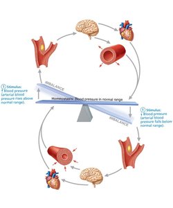
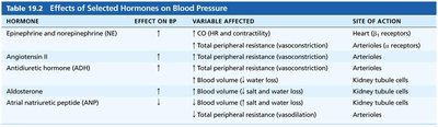
Short-term regulation involves neural reflexes (baroreceptors, chemoreceptors) and hormones (epinephrine, norepinephrine, angiotensin II, ADH, ANP) that alter vessel diameter and cardiac output.
Baroreceptor reflexes: Detect changes in blood pressure and adjust heart rate and vessel diameter accordingly.
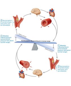
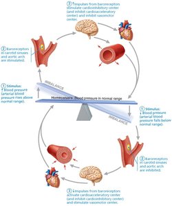
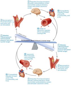
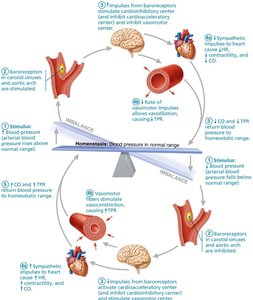
Hormonal effects: See table below for summary.
Hormone | Effect on BP | Variable Affected | Site of Action |
|---|---|---|---|
Epinephrine/Norepinephrine | ↑ | CO, TPR | Heart, arterioles |
Angiotensin II | ↑ | TPR | Arterioles |
ADH | ↑ | TPR, blood volume | Arterioles, kidneys |
Aldosterone | ↑ | Blood volume | Kidneys |
ANP | ↓ | Blood volume, TPR | Kidneys, arterioles |
Renal Regulation (Long-Term Control)
The kidneys regulate blood pressure by controlling blood volume through direct filtration and the renin-angiotensin-aldosterone system (RAAS). This system responds to low blood pressure by increasing blood volume and vasoconstriction.
Control of Blood Flow and Capillary Exchange
Intrinsic and Extrinsic Regulation
Blood flow to tissues is regulated by local (intrinsic) and systemic (extrinsic) mechanisms. Intrinsic controls (autoregulation) adjust blood flow to match tissue needs, while extrinsic controls (nervous and hormonal) redistribute blood during exercise or stress.
Capillary Exchange Mechanisms
Exchange of gases, nutrients, and wastes occurs across capillary walls by diffusion, fenestrations, and vesicular transport. Bulk flow of fluid is governed by hydrostatic and osmotic pressures, determining filtration and reabsorption.
Net filtration pressure (NFP):
Clinical Correlations
Homeostatic Imbalances
Hypertension: Chronic high blood pressure; major risk for heart failure, stroke, and kidney disease.
Hypotension: Abnormally low blood pressure; may cause inadequate tissue perfusion.
Circulatory shock: Inadequate blood flow to meet tissue needs; can result from blood loss, vasodilation, or heart failure.
Edema: Excess interstitial fluid due to increased capillary pressure, decreased plasma proteins, or lymphatic obstruction.
Circulatory Pathways
Pulmonary and Systemic Circulation
The vascular system consists of two main circuits:
Pulmonary circulation: Heart → lungs → heart (gas exchange)
Systemic circulation: Heart → body tissues → heart (nutrient/waste exchange)
Major Arteries and Veins
Major arteries and veins are named according to the regions, organs, or bones they serve or follow. Arteries tend to run deep, while veins can be both deep and superficial. The brain and digestive system have unique venous drainage systems (dural sinuses, hepatic portal system).
Developmental Aspects of Blood Vessels
Blood vessels develop from mesodermal blood islands in the embryo. Fetal circulation includes shunts to bypass nonfunctional lungs and liver, which close at birth. With aging, vessels may become less elastic, and vascular diseases such as atherosclerosis and varicose veins become more common.
