 Back
The Cardiovascular System: Blood Vessels – Structure, Function, and Physiology
Study Guide - Smart Notes
 Tailored notes based on your materials, expanded with key definitions, examples, and context.
Blood Vessel Structure and Function
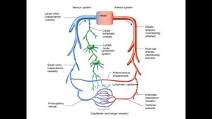
Overview of Blood Vessels
Blood vessels are dynamic organs of the cardiovascular system that form a closed circuit, transporting blood from the heart to body tissues and back. They are not static pipes but actively constrict, dilate, and adapt to physiological needs.
Arteries: Carry oxygenated blood away from the heart (except in pulmonary circulation).
Veins: Return oxygen-poor blood to the heart.
Capillaries: Microscopic vessels where exchange of gases, nutrients, and wastes occurs between blood and tissues.
Major Types of Blood Vessels
Arteries: Branch into smaller vessels as they move away from the heart.
Veins: Merge into larger vessels as they approach the heart.
Capillaries: Smallest vessels, site of exchange.
Structure of Blood Vessel Walls
Generalized Structure
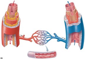
Most blood vessels (except capillaries) have three layers (tunics):
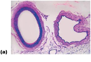
Tunica intima: Innermost layer, composed of endothelium (simple squamous epithelium) that minimizes friction.
Tunica media: Middle layer, primarily smooth muscle and elastin, responsible for vasoconstriction and vasodilation, regulated by the autonomic nervous system.
Tunica externa (adventitia): Outermost layer, composed of collagen fibers that protect, reinforce, and anchor the vessel. Large vessels contain vasa vasorum to nourish the vessel wall.
Vasoconstriction and Vasodilation
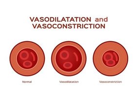
Changes in vessel diameter are crucial for regulating blood flow and pressure:
Vasoconstriction: Decrease in lumen diameter due to smooth muscle contraction.
Vasodilation: Increase in lumen diameter due to smooth muscle relaxation.
Types of Arteries
Classification by Size and Function
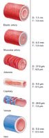
Elastic (Conducting) Arteries: Largest, near the heart (e.g., aorta), act as pressure reservoirs.
Muscular (Distributing) Arteries: Deliver blood to specific organs, thick tunica media, active in vasoconstriction.
Arterioles: Smallest arteries, control blood flow into capillary beds via constriction/dilation.
Clinical Applications: Arterial Diseases
Atherosclerosis and Arteriosclerosis
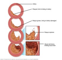
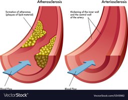
Atherosclerosis: Cholesterol plaques form in arterial walls, increasing blood pressure and risk of blockage.
Arteriosclerosis: Hardening and loss of elasticity of arteries, leading to intermittent blood flow.
Aneurysm
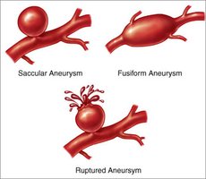
A bulge in a weakened artery wall, which can rupture and cause life-threatening bleeding.
Capillaries and Microcirculation
Structure and Function
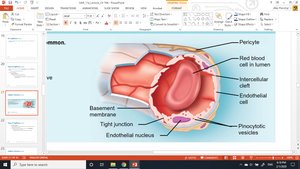
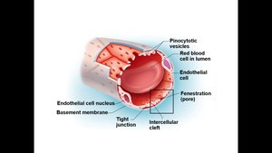
Capillaries are composed of a single layer of endothelial cells and a basement membrane, allowing efficient exchange between blood and tissues. Pericytes support capillary stability and repair.
Types of Capillaries
Continuous Capillaries: Most common, found in skin and muscles, form the blood-brain barrier.
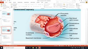
Fenestrated Capillaries: Have pores, found in kidneys, intestines, and endocrine glands for rapid exchange.
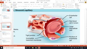
Sinusoidal Capillaries: Large, leaky, found in liver, bone marrow, spleen, and adrenal medulla.
Capillary Beds and Microcirculation
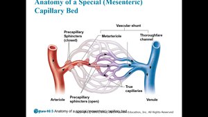
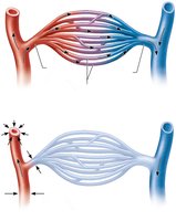
Capillary beds consist of networks of true capillaries and vascular shunts. Precapillary sphincters regulate blood flow through the bed, allowing for vasomotion (intermittent flow).
Venous System
Structure and Function
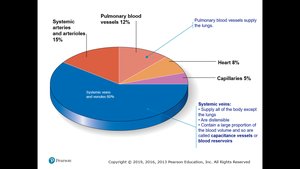
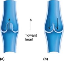
Veins return blood to the heart, have thinner walls and larger lumens than arteries, and contain valves to prevent backflow, especially in the limbs. Venules collect blood from capillaries and gradually merge into larger veins.
Venous Disorders
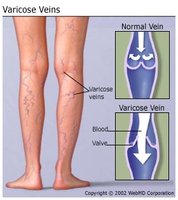
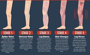
Varicose Veins: Result from weakened valves and walls, causing blood pooling and vein distension.
Hemorrhoids: Varicosities in the anal canal due to increased intra-abdominal pressure.
Physiology of Circulation
Blood Flow, Pressure, and Resistance
Blood Flow: Volume of blood moving through a vessel per unit time (mL/min).
Blood Pressure (BP): Force per unit area exerted by blood on vessel walls, measured in mm Hg.
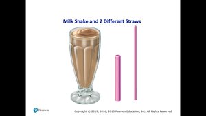
Resistance: Opposition to flow, mainly due to vessel diameter, blood viscosity, and vessel length.
Systemic Blood Pressure
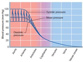
Highest in the aorta, declines through the vascular tree, lowest in the venae cavae.
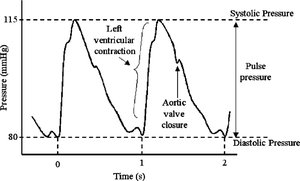
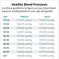
Systolic pressure: Peak pressure during ventricular contraction (~120 mm Hg).
Diastolic pressure: Lowest pressure during ventricular relaxation (~80 mm Hg).
Pulse pressure: Difference between systolic and diastolic pressures.
Measuring Blood Pressure
Measured indirectly using a sphygmomanometer and stethoscope (auscultatory method).
Systolic pressure: First appearance of Korotkoff sounds.
Diastolic pressure: Disappearance of sounds.
Venous Blood Pressure and Return
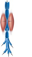
Venous pressure is low and steady; aided by muscular pump, respiratory pump, and sympathetic venoconstriction.
Regulation of Blood Pressure
Short-Term and Long-Term Controls
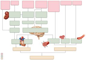
Short-term (Neural and Hormonal): Baroreceptors, chemoreceptors, and hormones (e.g., epinephrine, norepinephrine, angiotensin II, ADH, ANP) adjust vessel diameter and heart rate.
Long-term (Renal): Kidneys regulate blood volume by adjusting urine output.
Homeostatic Imbalances
Hypotension
Low blood pressure (systolic < 100 mm Hg), may be normal or indicate underlying disease.
Hypertension
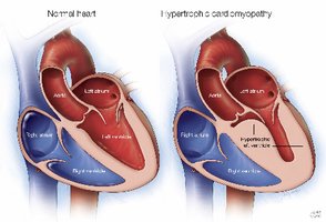
High blood pressure (>140/90 mm Hg), increases risk for heart failure, stroke, and kidney disease.
Primary hypertension: No identifiable cause, influenced by genetics, diet, obesity, age, diabetes, stress, and smoking.
Secondary hypertension: Due to identifiable disorders (e.g., renal disease, endocrine disorders).
Capillary Exchange and Bulk Flow
Mechanisms of Exchange
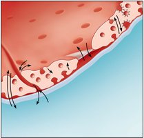
Diffusion: Lipid-soluble substances pass through endothelial membranes; water-soluble substances pass through clefts or fenestrations.
Active transport: Large molecules via vesicles or caveolae.
Bulk Flow and Fluid Movements
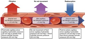
Driven by hydrostatic and osmotic pressures.
Net filtration at arterial end, net reabsorption at venous end.
Edema
Abnormal accumulation of interstitial fluid due to increased capillary hydrostatic pressure, decreased colloid osmotic pressure, or lymphatic obstruction.
Circulatory Shock
Hypovolemic shock: Due to large-scale blood loss.
Vascular shock: Extreme vasodilation (e.g., anaphylaxis).
Cardiogenic shock: Heart pump failure.
Blood Vessels of the Body
Major Circulatory Pathways
Pulmonary circulation: Heart to lungs and back.
Systemic circulation: Heart to all body tissues and back.
Arterial and Venous Pathways
Arteries branch from the aorta; veins converge toward the venae cavae.
Veins are more variable and interconnected than arteries.
Special venous systems: Dural sinuses (brain), hepatic portal system (digestive tract to liver).
Summary Table: Comparison of Arteries, Veins, and Capillaries
Feature | Arteries | Veins | Capillaries |
|---|---|---|---|
Wall Thickness | Thick | Thin | Very thin (one cell layer) |
Lumen Size | Narrow | Wide | Very narrow |
Valves | No | Yes (especially in limbs) | No |
Function | Carry blood away from heart | Carry blood toward heart | Exchange of substances |
Additional info: This guide covers the structure and function of blood vessels, their role in circulation, and clinical correlations relevant to Anatomy & Physiology II college courses.
