 Back
BackThe Cardiovascular System: Blood Vessels – Structure, Function, and Regulation
Study Guide - Smart Notes
 Tailored notes based on your materials, expanded with key definitions, examples, and context.
The Cardiovascular System: Blood Vessels
Introduction
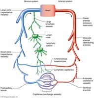
The cardiovascular system is responsible for the transport of blood throughout the body, ensuring the delivery of oxygen, nutrients, and the removal of waste products. Blood vessels form a complex network that begins and ends at the heart, working closely with the lymphatic system to maintain fluid balance and tissue health.
Blood Vessel Structure and Function
Types of Blood Vessels
Arteries: Carry blood away from the heart; typically oxygenated except in pulmonary circulation and fetal vessels.
Capillaries: Microscopic vessels that facilitate exchange between blood and tissues.
Veins: Return blood to the heart; typically deoxygenated except in pulmonary circulation and fetal vessels.
Layers of Blood Vessel Walls
Except for capillaries, all blood vessels have three layers (tunics):
Tunica intima: Innermost layer; endothelium provides a smooth, friction-reducing lining.
Tunica media: Middle layer; composed of smooth muscle and elastin, responsible for vasoconstriction and vasodilation.
Tunica externa (adventitia): Outermost layer; composed of collagen fibers, protects and anchors vessels, contains nerves and lymphatics.
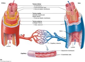
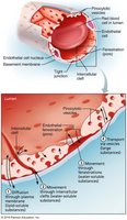
Capillary Structure
Capillaries consist of a single layer of endothelial cells and a sparse basal lamina, allowing efficient exchange of substances.
Types of Arteries
Elastic arteries: Large, thick-walled; act as pressure reservoirs (e.g., aorta).
Muscular arteries: Distribute blood to organs; thick tunica media, active in vasoconstriction.
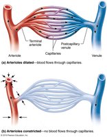
Arterioles: Smallest arteries; regulate blood flow into capillary beds via vasodilation/constriction.
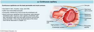
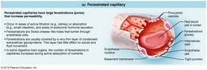
Types of Capillaries
Continuous capillaries: Least permeable; abundant in skin, muscles, lungs, CNS.
Fenestrated capillaries: Have pores (fenestrations) for increased permeability; found in kidneys, intestines, endocrine glands.
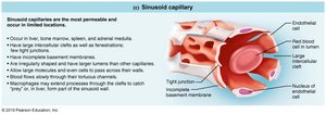
Sinusoidal capillaries: Most permeable; large clefts and lumens; found in liver, bone marrow, spleen, adrenal medulla.
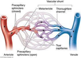
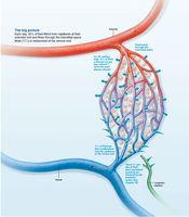
Capillary Beds
Capillary beds are networks of capillaries between arterioles and venules, facilitating exchange of gases, nutrients, and wastes. Flow is regulated by the diameter of arterioles and precapillary sphincters.
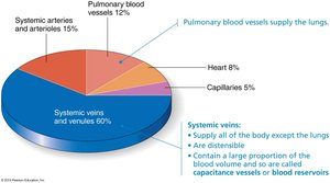
Veins
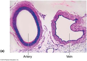
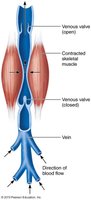
Veins have thinner walls and larger lumens than arteries, serving as blood reservoirs (capacitance vessels). They contain valves to prevent backflow, especially in limbs.
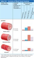
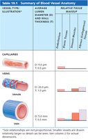
Summary Table: Blood Vessel Anatomy
Vessel Type | Diameter | Wall Thickness | Key Features |
|---|---|---|---|
Elastic artery | 1.0-2.5 cm | 1.0-1.5 mm | Pressure reservoir, high elastin |
Muscular artery | 0.3 mm-1.0 cm | 0.5-1.0 mm | Distributing, thick smooth muscle |
Arteriole | 10-300 μm | 6-10 μm | Resistance vessels |
Capillary | 8-10 μm | 0.5 μm | Exchange vessels |
Venule | 8-100 μm | 1.0 μm | Smallest veins |
Vein | 0.1 mm-2.5 cm | 0.5 mm | Capacitance vessels, valves |
Vascular Anastomoses
Vascular anastomoses are interconnections between blood vessels, providing alternate pathways for blood flow. They are common in joints, abdominal organs, brain, and heart.
Blood Flow, Blood Pressure, and Resistance
Definitions
Blood flow (F): Volume of blood moving through a vessel per unit time (ml/min).
Blood pressure (BP): Force per unit area exerted by blood on vessel walls (mm Hg).
Resistance (R): Opposition to flow, mainly due to vessel diameter, blood viscosity, and vessel length.
Relationship: $F = \frac{\Delta P}{R}$

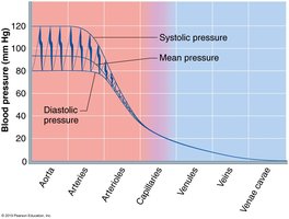
Systemic Blood Pressure
Blood pressure is highest in the aorta and decreases through the systemic circuit. Systolic pressure is the peak during ventricular contraction; diastolic is the lowest during relaxation. Mean arterial pressure (MAP) is the average pressure driving blood to tissues.
MAP calculation: $MAP = \text{Diastolic Pressure} + \frac{1}{3}(\text{Pulse Pressure})$
Measuring Blood Pressure
Measured indirectly using a sphygmomanometer and stethoscope (auscultatory method).
Systolic: pressure when sounds first heard (<120 mm Hg).
Diastolic: pressure when sounds disappear (<80 mm Hg).
Venous Blood Pressure and Return
Venous pressure is low and aided by muscular pump, respiratory pump, and sympathetic venoconstriction.
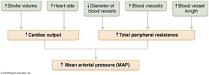
Regulation of Blood Pressure
Blood pressure is regulated by cardiac output (CO), peripheral resistance (PR), and blood volume. Regulation involves neural, hormonal, and renal mechanisms.
Neural Controls
Baroreceptor reflexes: Detect changes in pressure and adjust vessel diameter and heart rate.
Chemoreceptor reflexes: Respond to CO2, pH, and O2 changes, influencing CO and vasoconstriction.
Higher brain centers: Hypothalamus and cortex can modify BP during stress, exercise, or temperature changes.
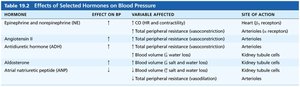
Hormonal Controls
Epinephrine/norepinephrine: Increase CO and vasoconstriction.
Angiotensin II: Potent vasoconstrictor.
ADH: Increases blood volume and vasoconstriction at high levels.
Atrial natriuretic peptide: Decreases blood volume and BP.
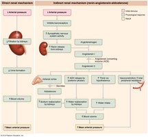
Renal Regulation
Direct mechanism: Alters urine output to regulate blood volume.
Indirect mechanism: Renin-angiotensin-aldosterone system increases BP via vasoconstriction and water retention.

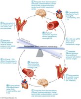
Homeostatic Imbalances
Hypertension: Sustained high BP (>140/90 mm Hg); risk factors include genetics, diet, obesity, age, and stress.
Hypotension: Low BP (<90/60 mm Hg); may indicate underlying health issues.
Circulatory shock: Inadequate blood flow to meet tissue needs; can be hypovolemic, vascular, or cardiogenic in origin.
Control of Blood Flow and Capillary Exchange
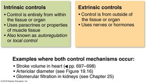
Intrinsic vs. Extrinsic Controls
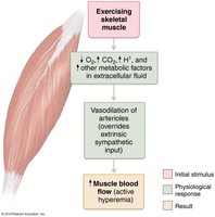
Intrinsic (autoregulation): Local regulation to meet tissue needs (metabolic and myogenic mechanisms).
Extrinsic: Nervous and hormonal regulation for systemic needs.
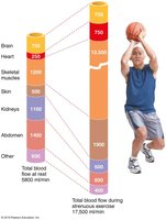
Blood Flow in Special Areas
Skeletal muscle: Increases greatly during exercise (active hyperemia).
Brain: Maintains constant flow; sensitive to CO2 and pH.
Skin: Regulates temperature and acts as a blood reservoir.
Lungs: Low pressure, unique autoregulation (low O2 causes vasoconstriction).
Heart: Flow increases with activity; high O2 extraction at rest.
Capillary Exchange and Bulk Flow
Mechanisms of Exchange
Diffusion through membranes (lipid-soluble substances).
Passage through intercellular clefts or fenestrations (water-soluble substances).
Active transport via vesicles (large molecules).

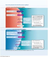
Bulk Flow and Fluid Movements
Bulk flow is the movement of fluid across capillary walls, driven by hydrostatic and osmotic pressures. Net filtration pressure (NFP) determines the direction of fluid movement:
$NFP = (HP_c + OP_{if}) - (HP_{if} + OP_c)$
Filtration occurs at the arterial end; reabsorption at the venous end.
Excess fluid is returned to the blood via the lymphatic system.


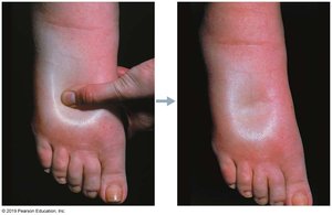
Edema
Edema is the abnormal accumulation of interstitial fluid, caused by increased capillary hydrostatic pressure, decreased plasma protein concentration, or impaired lymphatic drainage.
Circulatory Pathways
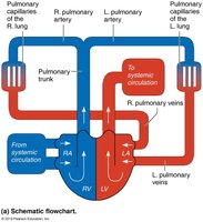
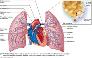
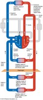
Pulmonary and Systemic Circulation
Pulmonary circuit: Short loop from heart to lungs and back; oxygenates blood.
Systemic circuit: Long loop to all body tissues and back; delivers oxygen and nutrients.
Developmental Aspects of Blood Vessels
Blood vessels develop from mesodermal blood islands in the embryo.
Fetal shunts (foramen ovale, ductus arteriosus, ductus venosus) bypass nonfunctional organs.
With aging, vessels may develop varicosities, atherosclerosis, and increased blood pressure.
Additional info: This guide covers the structure, function, and regulation of blood vessels, including clinical correlations and developmental aspects, as outlined in a typical college-level Human Anatomy & Physiology course.
