 Back
BackThe Cardiovascular System: Blood Vessels – Structure, Function, and Regulation
Study Guide - Smart Notes
 Tailored notes based on your materials, expanded with key definitions, examples, and context.
The Cardiovascular System: Blood Vessels
Introduction
The cardiovascular system is essential for transporting blood throughout the body, delivering oxygen and nutrients, and removing waste products. Blood vessels form a complex network that ensures efficient circulation and tissue perfusion. This chapter focuses on the structure, function, and regulation of blood vessels, as well as clinical aspects related to vascular health.
Blood Vessel Structure and Function
Types of Blood Vessels
Arteries: Carry blood away from the heart; usually oxygenated except in pulmonary and fetal circulation.
Capillaries: Microscopic vessels that directly serve tissue cells and facilitate exchange of substances.
Veins: Return blood to the heart; usually deoxygenated except in pulmonary and fetal circulation.
General Structure of Vessel Walls
Except for capillaries, all blood vessels have three layers (tunics):
Tunica intima: Innermost layer; endothelium provides a smooth, friction-reducing lining.
Tunica media: Middle layer; composed of smooth muscle and elastin, responsible for vasoconstriction and vasodilation.
Tunica externa (adventitia): Outermost layer; composed of collagen fibers, protects and anchors vessels.
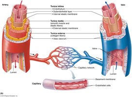
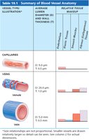
Capillary Structure
Capillaries consist of a single layer of endothelial cells and a sparse basal lamina, allowing efficient exchange between blood and tissues.
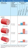
Types of Arteries
Elastic arteries: Large, thick-walled; act as pressure reservoirs (e.g., aorta).
Muscular arteries: Distribute blood to organs; thick tunica media, active in vasoconstriction.
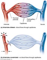
Arterioles: Smallest arteries; regulate blood flow into capillary beds.
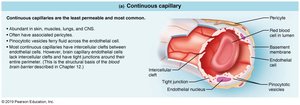
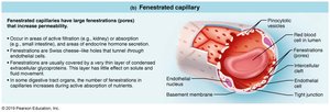
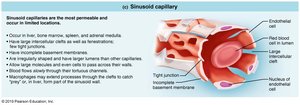
Types of Capillaries
Continuous capillaries: Least permeable; abundant in skin, muscles, lungs, and CNS.
Fenestrated capillaries: Have pores (fenestrations) for increased permeability; found in kidneys, intestines, endocrine glands.
Sinusoidal capillaries: Most permeable; large clefts and lumens; found in liver, bone marrow, spleen, adrenal medulla.
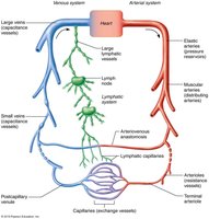
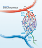
Capillary Beds
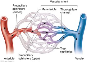
Capillary beds are networks between arterioles and venules, regulating microcirculation and exchange of substances.
Special features include vascular shunts (direct arteriole-to-venule channels) and precapillary sphincters (regulate flow into true capillaries).
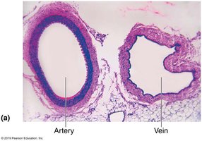
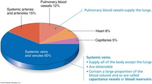
Veins
Veins have thinner walls and larger lumens than arteries, serving as blood reservoirs. They contain valves to prevent backflow, especially in limbs.
Summary Table: Blood Vessel Anatomy
Vessel Type | Diameter | Wall Thickness | Key Features |
|---|---|---|---|
Elastic artery | 1.0–2.5 cm | 1.0–1.5 mm | Pressure reservoir, high elastin |
Muscular artery | 0.3 mm–1.0 cm | 0.5–1.0 mm | Thick tunica media, active in vasoconstriction |
Arteriole | 10–300 μm | 6–10 μm | Regulate flow into capillaries |
Capillary | 8–10 μm | 0.5 μm | Exchange vessels, thin wall |
Venule | 8–100 μm | 1.0 μm | Collect blood from capillaries |
Vein | 0.1 mm–2.5 cm | 0.5 mm | Valves, capacitance vessels |
Vascular Anastomoses
Vascular anastomoses are interconnections between blood vessels, providing alternate pathways for blood flow. They are common in joints, abdominal organs, brain, and heart.
Blood Flow, Pressure, and Resistance
Definitions
Blood flow (F): Volume of blood moving through a vessel per unit time (ml/min).
Blood pressure (BP): Force per unit area exerted by blood on vessel walls (mm Hg).
Resistance (R): Opposition to flow, mainly due to vessel diameter, blood viscosity, and vessel length.
Relationship: $F = \frac{\Delta P}{R}$

Systemic Blood Pressure
Blood pressure is highest in the aorta and decreases through the systemic circuit. Arterial pressure is pulsatile near the heart and becomes steady in capillaries and veins.
Key Terms
Systolic pressure: Peak pressure during ventricular contraction (~120 mm Hg).
Diastolic pressure: Lowest pressure during ventricular relaxation (~80 mm Hg).
Pulse pressure: Difference between systolic and diastolic pressure.
Mean arterial pressure (MAP): Average pressure propelling blood to tissues. $MAP = \text{Diastolic Pressure} + \frac{1}{3}(\text{Pulse Pressure})$
Venous Return Mechanisms
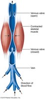
Muscular pump: Skeletal muscle contractions help push blood toward the heart.
Respiratory pump: Pressure changes during breathing aid venous return.
Venous valves: Prevent backflow of blood.
Regulation of Blood Pressure
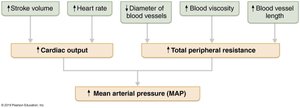
Main Factors
Cardiac output (CO)
Peripheral resistance (PR)
Blood volume
Blood pressure varies directly with CO, PR, and blood volume.
Short-Term Regulation
Neural controls: Baroreceptor and chemoreceptor reflexes, cardiovascular centers in the medulla.
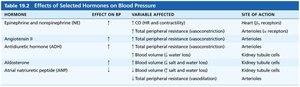
Hormonal controls: Epinephrine, norepinephrine, angiotensin II, ADH, and atrial natriuretic peptide (ANP).
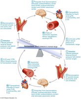
Long-Term Regulation
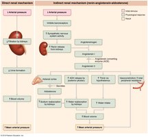
Renal mechanisms: Direct (altering urine output) and indirect (renin-angiotensin-aldosterone system) control blood volume and pressure.

Homeostatic Imbalances
Hypertension: Sustained high blood pressure (≥140/90 mm Hg); risk factor for heart failure, stroke, and kidney disease.
Hypotension: Low blood pressure (≤90/60 mm Hg); may cause inadequate tissue perfusion.
Circulatory shock: Inadequate blood flow to meet tissue needs; can be hypovolemic, vascular, or cardiogenic.
Control of Blood Flow and Capillary Exchange
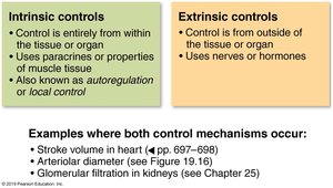
Intrinsic vs. Extrinsic Controls
Intrinsic (autoregulation): Local regulation to meet tissue needs (metabolic and myogenic mechanisms).
Extrinsic: Nervous and hormonal regulation for overall blood distribution.
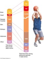
Blood Flow in Special Areas
Skeletal muscle: Increases during exercise (active hyperemia).
Brain: Maintains constant flow; sensitive to CO2 and pH changes.
Skin: Regulates temperature and acts as a blood reservoir.
Lungs: Low pressure, unique autoregulation (low O2 causes vasoconstriction).
Heart: Coronary circulation increases with activity.
Capillary Exchange and Bulk Flow
Mechanisms of Exchange
Diffusion through membranes (lipid-soluble substances)
Passage through intercellular clefts (water-soluble substances)
Passage through fenestrations
Active transport via vesicles
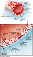
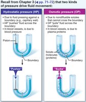
Bulk Flow and Fluid Movements
Bulk flow is the movement of fluid across capillary walls, driven by hydrostatic and osmotic pressures. Net filtration pressure (NFP) determines the direction of fluid movement:
$NFP = (HP_c + OP_{if}) - (HP_{if} + OP_c)$
Filtration occurs at the arterial end; reabsorption at the venous end.
Excess fluid is returned to the blood via the lymphatic system.
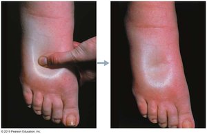
Clinical Correlation: Edema
Edema is the abnormal accumulation of interstitial fluid, caused by increased capillary hydrostatic pressure, decreased plasma protein concentration, or lymphatic obstruction.
Circulatory Pathways
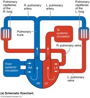
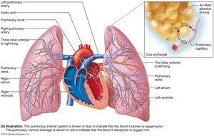
Pulmonary and Systemic Circulation
Pulmonary circuit: Short loop from heart to lungs and back; oxygenates blood.
Systemic circuit: Long loop from heart to all body tissues and back; delivers oxygen and nutrients.
Major Arteries and Veins
Arteries and veins are named according to body regions, organs, or bones they serve or follow.
Venous pathways are more interconnected and variable than arterial pathways.
Developmental Aspects
Blood vessels develop from mesodermal blood islands in the embryo.
Fetal shunts bypass nonfunctional lungs and liver.
With aging, vessels may develop varicosities, atherosclerosis, and increased blood pressure.
