 Back
The Cardiovascular System: Blood Vessels – Structured Study Notes
Study Guide - Smart Notes
 Tailored notes based on your materials, expanded with key definitions, examples, and context.
The Cardiovascular System: Blood Vessels
Overview of Blood Vessels
The cardiovascular system consists of a network of blood vessels that transport blood throughout the body. These vessels are classified as arteries, capillaries, and veins, each serving distinct functions in circulation.
Arteries: Carry blood away from the heart; most are oxygenated except for pulmonary arteries and fetal umbilical arteries.
Capillaries: Microscopic vessels that directly serve tissue cells, facilitating exchange of gases, nutrients, and wastes.
Veins: Return blood to the heart; most carry deoxygenated blood except for pulmonary veins and fetal umbilical veins.
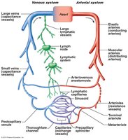
Structure of Blood Vessel Walls
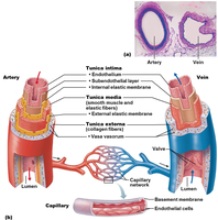
Blood vessel walls are composed of three layers, or tunics, which provide structural integrity and regulate vessel function. Capillaries have a simpler structure, consisting mainly of endothelium.
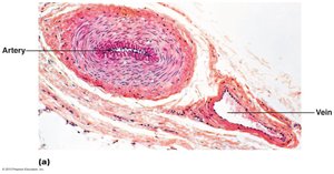
Tunica intima: Innermost layer; consists of endothelium lining the lumen and, in larger vessels, a subendothelial layer and basement membrane.
Tunica media: Middle layer; composed of smooth muscle and elastin, responsible for vasoconstriction and vasodilation.
Tunica externa (adventitia): Outermost layer; made of collagen fibers, provides protection and reinforcement. Large vessels contain vasa vasorum to nourish the external layer.
Lumen: Central blood-containing space.
Types of Arteries
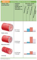
Arteries are classified based on their size and function. Elastic arteries act as pressure reservoirs, while muscular arteries distribute blood to organs, and arterioles regulate blood flow into capillary beds.
Elastic (Conducting) Arteries: Large, thick-walled arteries (e.g., aorta) with elastin in all three tunics; expand and recoil to maintain blood pressure.
Muscular (Distributing) Arteries: Medium-sized arteries with more smooth muscle; actively regulate blood flow via vasoconstriction.
Arterioles: Smallest arteries; control flow into capillary beds through vasodilation and vasoconstriction.
Capillaries: Structure and Function
Capillaries are the smallest blood vessels, consisting of a thin tunica intima. Their structure allows for efficient exchange of substances between blood and tissues.
Endothelium: Single layer of cells lining the capillary.
Pericytes: Contractile cells that stabilize capillary walls and regulate permeability.
Size: Only large enough for a single red blood cell to pass at a time.
Distribution: Found in all tissues except cartilage, epithelia, cornea, and lens.
Function: Exchange of gases, nutrients, wastes, and hormones.
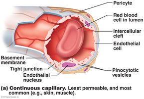
Types of Capillaries
Capillaries are classified into three structural types based on their permeability and location.
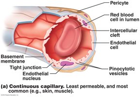
Continuous Capillaries: Most common, least permeable; found in skin and muscles. Tight junctions connect endothelial cells, but intercellular clefts allow passage of fluids and small solutes. In the brain, tight junctions form the blood-brain barrier.
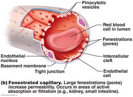
Fenestrated Capillaries: Endothelial cells contain pores (fenestrations); more permeable, found in areas of absorption or filtration (e.g., kidneys, small intestine).
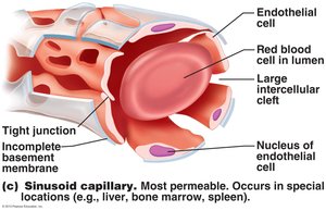
Sinusoidal Capillaries (Sinusoids): Most permeable; large intercellular clefts and incomplete basement membrane. Found in liver, bone marrow, and spleen; allow passage of large molecules and cells.
Capillary Beds and Microcirculation
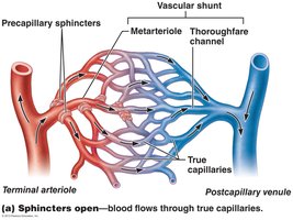
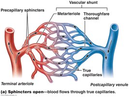
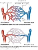
Capillary beds are networks of capillaries that facilitate microcirculation between arterioles and venules. Blood flow through these beds is regulated by precapillary sphincters.
Vascular shunt: Direct connection between terminal arteriole and postcapillary venule (metarteriole-thoroughfare channel).
True capillaries: Branch off the metarteriole; 10 to 100 exchange vessels per bed.
Precapillary sphincters: Regulate blood flow into true capillaries based on local chemical conditions and vasomotor nerves.
Venules and Veins
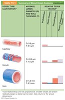
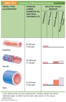
Venules are formed when capillary beds unite and are very porous, allowing fluids and white blood cells to enter tissues. Veins are formed when venules converge and serve as blood reservoirs.
Venules: Smallest veins; postcapillary venules consist of endothelium and a few pericytes. Larger venules have smooth muscle layers.
Veins: Thinner walls and larger lumens than arteries; contain up to 65% of blood supply. Adaptations include valves to prevent backflow and large-diameter lumens for low resistance.
Venous sinuses: Flattened veins with extremely thin walls (e.g., coronary sinus, dural sinuses).
Vascular Anastomoses
Anastomoses are interconnections between blood vessels that provide alternate pathways for blood flow. They are common in joints, abdominal organs, brain, and heart.
Arterial anastomoses: Provide collateral channels to a region.
Venous anastomoses: More common; blockage rarely leads to tissue death.
Arteriovenous anastomoses: Vascular shunts in capillaries.
Physiology of Circulation
Blood Flow, Blood Pressure, and Resistance
Blood flow, blood pressure, and resistance are key concepts in circulatory physiology. Blood flow is the volume of blood moving through vessels, blood pressure is the force exerted by blood on vessel walls, and resistance is the opposition to flow.
Blood flow (F): Measured in ml/min; equivalent to cardiac output (CO) for the entire vascular system.
Blood pressure (BP): Expressed in mm Hg; measured as systemic arterial BP in large arteries near the heart.
Resistance (R): Determined by blood viscosity, vessel length, and vessel diameter.
Key Equation:
Where is blood flow, is the pressure gradient, and is resistance.
Factors Affecting Resistance
Resistance is influenced by blood viscosity, vessel length, and vessel diameter. Vessel diameter is the most variable and has the greatest effect on resistance.
Blood viscosity: "Stickiness" due to formed elements and plasma proteins.
Vessel length: Longer vessels increase resistance.
Vessel diameter: Resistance varies inversely with the fourth power of radius.
Example: If radius is doubled, resistance is 1/16 as much; if halved, resistance increases 16-fold.
Systemic Blood Pressure
Blood pressure is highest in the aorta and declines throughout the circulatory pathway, reaching its lowest point in the right atrium. The steepest drop occurs in arterioles, which offer the greatest resistance.
Arterial Blood Pressure
Arterial blood pressure is affected by the elasticity of arteries and the volume of blood forced into them. It is pulsatile near the heart.
Systolic pressure: Pressure during ventricular contraction.
Diastolic pressure: Lowest arterial pressure.
Pulse pressure: Difference between systolic and diastolic pressure.
Mean arterial pressure (MAP): Pressure that propels blood to tissues.
MAP Equation:
Example: For BP 120/80 mmHg: mmHg
Capillary and Venous Blood Pressure
Capillary blood pressure is low (35–15 mm Hg) to prevent rupture and allow exchange. Venous blood pressure is even lower, aided by mechanisms such as the respiratory and muscular pumps, and sympathetic vasoconstriction.
Maintaining Blood Pressure
Blood pressure is regulated by cardiac output, peripheral resistance, and blood volume. Changes in one variable are compensated by changes in others to maintain homeostasis.
Cardiac output (CO): Determined by venous return, neural, and hormonal controls.
Peripheral resistance (PR): Influenced by vessel diameter and blood viscosity.
Blood volume: Regulated by kidneys.
Key Equation:
Control of Blood Pressure
Blood pressure is controlled by short-term neural and hormonal mechanisms, and long-term renal regulation.
Short-term: Neural (baroreceptor and chemoreceptor reflexes) and hormonal controls.
Long-term: Renal regulation (direct and indirect mechanisms).
Short-Term Neural Controls
Neural controls operate via reflex arcs involving baroreceptors, chemoreceptors, and higher brain centers. The vasomotor center in the medulla oversees vessel diameter.
Baroreceptors: Located in carotid sinuses, aortic arch, and large arteries; respond to changes in pressure.
Chemoreceptors: Located in carotid sinus, aortic arch, and large arteries; respond to changes in CO2, pH, and O2.
Higher brain centers: Cortex and hypothalamus can modify BP via medullary centers.
Short-Term Hormonal Controls
Hormones such as norepinephrine, epinephrine, angiotensin II, ADH, and ANP regulate blood pressure by affecting vessel diameter and blood volume.
Norepinephrine/Epinephrine: Cause vasoconstriction and increase cardiac output.
Angiotensin II: Potent vasoconstrictor; stimulates aldosterone and ADH release.
ADH: Causes vasoconstriction in cases of low BP.
ANP: Causes vasodilation and decreases blood volume.
Long-Term Renal Regulation
Kidneys regulate blood pressure by controlling blood volume through direct and indirect mechanisms.
Direct mechanism: Increased BP or blood volume leads to increased urine output, reducing BP; decreased BP leads to water conservation, raising BP.
Indirect mechanism: Renin-angiotensin system increases BP via vasoconstriction and sodium/water retention.
Renin-Angiotensin Mechanism:
Clinical Aspects and Homeostatic Imbalances
Measuring Blood Pressure
Blood pressure is measured using a sphygmomanometer and auscultatory method. Systolic and diastolic pressures are determined by listening for Korotkoff sounds.
Variations and Alterations in Blood Pressure
Hypotension: Systolic pressure below 100 mm Hg; may be orthostatic, chronic, or acute.
Circulatory shock: Inadequate blood flow; includes hypovolemic, vascular, and cardiogenic shock.
Hypertension: Sustained BP of 140/90 mm Hg or higher; primary (essential) or secondary.
Blood Flow Through Body Tissues
Autoregulation
Autoregulation is the automatic adjustment of blood flow to tissues based on their needs. It is controlled by metabolic and myogenic mechanisms.
Metabolic controls: Vasodilation in response to low O2, high CO2, H+, K+, adenosine, and prostaglandins.
Myogenic controls: Vascular smooth muscle responds to changes in pressure; increased stretch causes vasoconstriction, reduced stretch causes vasodilation.
Angiogenesis: Long-term increase in vessel number or size when nutrient requirements are unmet.
Blood Flow in Specific Organs
Skeletal muscles: Blood flow increases with activity; local controls override sympathetic vasoconstriction.
Brain: Constant blood flow; sensitive to pH and CO2 changes.
Skin: Regulates temperature and serves as a blood reservoir.
Lungs: Low resistance and pressure; vasoconstriction occurs in low O2 areas.
Heart: Blood flow increases during exercise; myogenic and metabolic controls predominate.
Capillary Exchange and Fluid Movements
Capillary Exchange
Exchange of gases, nutrients, and wastes occurs via diffusion, with lipid-soluble molecules passing through membranes, water-soluble solutes through clefts and fenestrations, and large molecules via vesicular transport.
Bulk Flow and Fluid Movements
Bulk flow determines fluid volumes in blood and interstitial space, governed by hydrostatic and colloid osmotic pressures.
Hydrostatic pressure (HPc): Forces fluids out of capillaries; higher at arterial end.
Colloid osmotic pressure (OPc): Draws water into capillaries due to plasma proteins.
Net Filtration Pressure (NFP): Determines direction and amount of fluid movement.
NFP Equation:
At the arterial end, hydrostatic forces dominate; at the venous end, osmotic forces dominate. Excess fluid is returned via the lymphatic system.
