Back
BackThe Cardiovascular System: General Circulation and the Heart
Study Guide - Smart Notes
 Tailored notes based on your materials, expanded with key definitions, examples, and context.
General Circulatory System
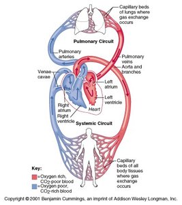
Overview of the Cardiovascular System
The cardiovascular system is a closed network of blood vessels responsible for transporting blood throughout the body. It consists of two main circuits: the systemic circuit, which delivers oxygenated blood to body tissues, and the pulmonary circuit, which transports deoxygenated blood to the lungs for oxygenation. Arteries carry blood away from the heart, while veins return blood to the heart.
Systemic Circuit: Delivers oxygen-rich blood from the left side of the heart to the body and returns oxygen-poor blood to the right side of the heart.
Pulmonary Circuit: Carries oxygen-poor blood from the right side of the heart to the lungs and returns oxygen-rich blood to the left side of the heart.
Arteries: Vessels that transport blood away from the heart.
Veins: Vessels that transport blood toward the heart.
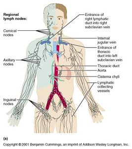
Lymphvascular System
The lymphvascular system is a network of vessels that collect interstitial fluid (lymph) from tissues and return it to the bloodstream. Lymph is filtered through lymph nodes before re-entering circulation, playing a key role in immune defense and fluid balance.
Lymphatic Vessels: Begin as blind-ended tubes in tissues, collecting excess interstitial fluid.
Lymph Nodes: Filter lymph, removing pathogens and debris before lymph rejoins the blood.
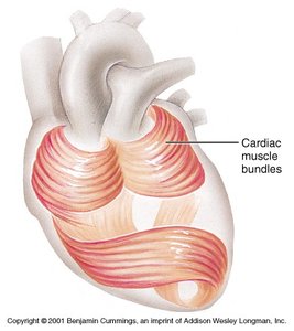
Heart as a Dual Pump
Cardiac Muscle Structure and Function
The heart functions as a dual pump, with the right side pumping blood to the lungs (pulmonary circuit) and the left side pumping blood to the body (systemic circuit). Cardiac muscle fibers are arranged in spiral bundles (whorls) that efficiently squeeze blood through the chambers.
Four Chambers: Two atria (upper chambers) and two ventricles (lower chambers).
Muscle Arrangement: Spiral bundles enhance the efficiency of blood ejection.
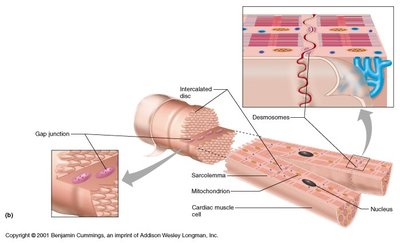
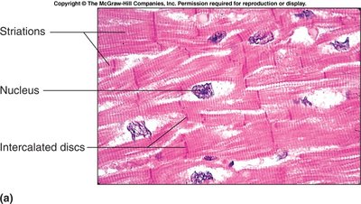
Cardiac Muscle Cells
Cardiac muscle cells (cardiomyocytes) are specialized for continuous rhythmic contraction. They are connected by intercalated discs, which contain gap junctions and desmosomes, allowing for synchronized contraction and mechanical stability.
Intercalated Discs: Specialized connections between cells that facilitate electrical and mechanical coupling.
Gap Junctions: Allow ions to pass directly between cells, enabling rapid electrical communication.
Desmosomes: Provide strong adhesion between cells during contraction.
Cardiac Muscle Depolarization and Action Potentials
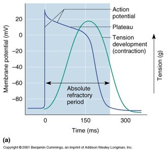
Phases of Cardiac Action Potential
Cardiac muscle contraction is initiated by an action potential, which has distinct phases: rapid depolarization, plateau, and repolarization. The plateau phase is unique to cardiac muscle and is responsible for the prolonged contraction necessary for effective blood ejection.
Depolarization: Rapid influx of Na+ ions.
Plateau Phase: Sustained influx of Ca2+ ions, maintaining depolarization.
Repolarization: Efflux of K+ ions restores resting membrane potential.
Absolute Refractory Period: Period during which the cell cannot be re-excited, preventing tetanus.
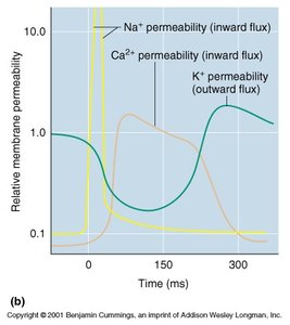
Ion Conductance During Depolarization
The conductance of sodium, calcium, and potassium ions changes during the cardiac action potential, producing the characteristic phases of depolarization, plateau, and repolarization.
Na+ Permeability: Increases rapidly during depolarization.
Ca2+ Permeability: Increases during the plateau phase, sustaining contraction.
K+ Permeability: Increases during repolarization, restoring resting potential.
Heart Development and Fetal Circulation
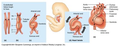
Embryonic Heart Development
The heart develops from a simple tube that undergoes folding and partitioning to form a four-chambered organ. This process ensures the separation of oxygenated and deoxygenated blood.
Fusion of Endothelial Tubes: Forms the primitive heart tube.
Heart Looping: Establishes the basic shape and orientation of the heart.
Chamber Formation: Septa develop to separate atria and ventricles.
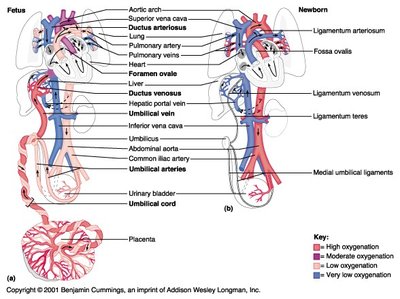
Fetal Circulation
Fetal circulation includes unique structures that bypass the nonfunctional fetal lungs, such as the foramen ovale and ductus arteriosus. After birth, these structures close as the newborn's lungs become functional.
Foramen Ovale: Opening between right and left atria, allowing blood to bypass the lungs.
Ductus Arteriosus: Vessel connecting pulmonary artery to aorta, diverting blood from the lungs.
Umbilical Vessels: Carry oxygenated blood from the placenta to the fetus and return deoxygenated blood to the placenta.
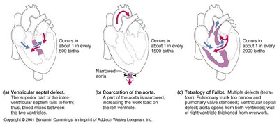
Selected Heart Defects
Congenital heart defects are structural abnormalities present at birth. Common examples include ventricular septal defect, coarctation of the aorta, and tetralogy of Fallot.
Ventricular Septal Defect: Opening in the interventricular septum, allowing blood to mix between ventricles.
Coarctation of the Aorta: Narrowing of the aorta, increasing workload on the left ventricle.
Tetralogy of Fallot: Combination of four defects, including pulmonary stenosis and ventricular septal defect.