 Back
BackThe Cardiovascular System: Heart Anatomy and Physiology
Study Guide - Smart Notes
 Tailored notes based on your materials, expanded with key definitions, examples, and context.
Heart Anatomy and Physiology
Overview of the Cardiovascular System
The heart is a muscular organ that functions as the central component of the cardiovascular system, pumping blood throughout the body via two main circuits: the pulmonary and systemic circuits. It ensures the delivery of oxygen and nutrients to tissues and the removal of carbon dioxide and metabolic wastes.
Pulmonary circuit: Right side of the heart pumps oxygen-poor blood to the lungs for gas exchange.
Systemic circuit: Left side of the heart pumps oxygen-rich blood to the rest of the body.
Size, Location, and Orientation of the Heart
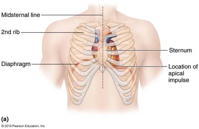
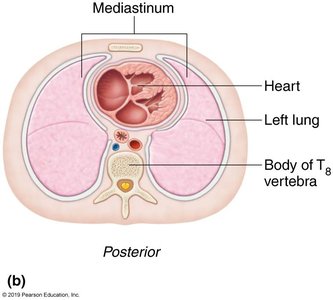
The heart is approximately the size of a fist and weighs less than one pound. It is located in the mediastinum, between the second rib and fifth intercostal space, resting on the superior surface of the diaphragm. Two-thirds of the heart lies to the left of the midsternal line, and its apex points toward the left hip.
Base: Leans toward the right shoulder.
Apex: Points toward the left hip; the apical impulse can be palpated between the fifth and sixth ribs, just below the left nipple.
Coverings of the Heart
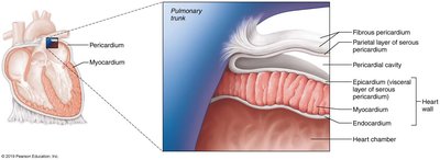
The heart is enclosed in a double-walled sac called the pericardium:
Fibrous pericardium: Tough, dense connective tissue that protects, anchors, and prevents overfilling.
Serous pericardium: Thin, two-layered membrane (parietal and visceral layers) separated by the pericardial cavity, which contains fluid to reduce friction.
Clinical Note: Pericarditis
Inflammation of the pericardium, causing friction rub and possibly cardiac tamponade (compression of the heart by excess fluid).
Layers of the Heart Wall
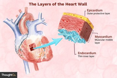
The heart wall consists of three layers:
Epicardium: Outer layer, also the visceral layer of the serous pericardium.
Myocardium: Middle, muscular layer composed of cardiac muscle bundles arranged in circular and spiral patterns for effective contraction.
Endocardium: Inner layer, continuous with the endothelial lining of blood vessels, lines heart chambers and covers valves.
Chambers and Associated Great Vessels
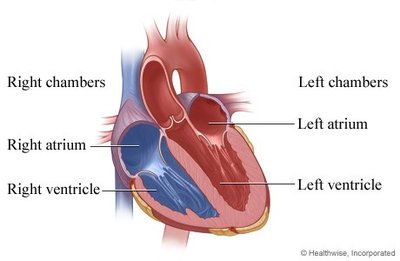
The heart has four chambers:
Atria (right and left): Superior, thin-walled chambers that receive blood returning to the heart.
Ventricles (right and left): Inferior, thick-walled chambers that discharge blood from the heart.
Right atrium: Receives deoxygenated blood from the superior and inferior vena cava and coronary sinus.
Left atrium: Receives oxygenated blood from the four pulmonary veins.
Right ventricle: Pumps blood into the pulmonary trunk (to the lungs).
Left ventricle: Pumps blood into the aorta (to the systemic circulation).
Surface Features and Internal Structures
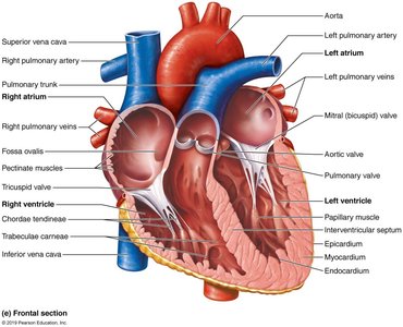
Coronary sulcus (atrioventricular groove): Encircles the junction of the atria and ventricles.
Anterior and posterior interventricular sulci: Mark the position of the interventricular septum.
Pectinate muscles: Ridges in the atria, especially prominent in the right atrium.
Trabeculae carneae: Irregular muscle ridges in the ventricles.
Papillary muscles: Project into ventricular cavities and anchor chordae tendineae.
Heart Valves
Heart valves ensure unidirectional blood flow through the heart and open/close in response to pressure changes.
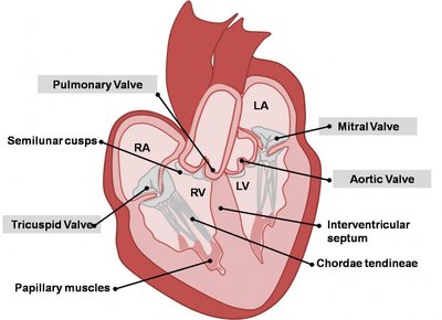
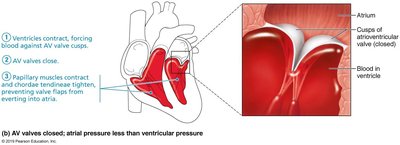
Atrioventricular (AV) valves: Between atria and ventricles (tricuspid on right, mitral/bicuspid on left).
Semilunar (SL) valves: Between ventricles and major arteries (pulmonary and aortic valves).
Function of AV Valves
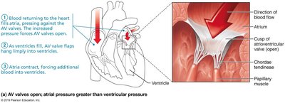
Prevent backflow into atria when ventricles contract.
Chordae tendineae anchor valve cusps to papillary muscles.
Function of SL Valves
Prevent backflow from arteries into ventricles.
Open and close in response to pressure changes.
Clinical Note: Valve Disorders
Incompetent valve: Blood backflows, heart repumps same blood.
Valvular stenosis: Stiff valve flaps constrict opening, requiring more force to pump blood.
Pathway of Blood Through the Heart
Blood flows through the heart in a specific sequence, ensuring separation of oxygenated and deoxygenated blood:
Deoxygenated blood enters right atrium via superior/inferior vena cava and coronary sinus.
Right atrium → tricuspid valve → right ventricle → pulmonary semilunar valve → pulmonary trunk → pulmonary arteries → lungs.
Oxygenated blood returns via pulmonary veins → left atrium → mitral valve → left ventricle → aortic semilunar valve → aorta → systemic circulation.
Ventricular Differences
Left ventricle has thicker walls and pumps with greater pressure to overcome systemic resistance.
Right ventricle pumps to the lungs, a shorter, lower-pressure circuit.
Coronary Circulation
The heart muscle (myocardium) receives its own blood supply via the coronary arteries and veins.
Coronary arteries: Arise from the base of the aorta; left and right coronary arteries branch to supply the heart.
Coronary veins: Collect deoxygenated blood from the myocardium and empty into the coronary sinus, which drains into the right atrium.
Clinical Note: Coronary Disorders
Angina pectoris: Chest pain due to transient ischemia of the myocardium.
Myocardial infarction (heart attack): Prolonged coronary blockage leading to cell death and scar tissue formation.
Microscopic Anatomy of Cardiac Muscle
Cardiac muscle cells are striated, short, branched, and interconnected. They contain one or two central nuclei and numerous mitochondria for resistance to fatigue. The cells are connected by intercalated discs, which contain desmosomes (mechanical connection) and gap junctions (electrical connection), allowing the heart to function as a coordinated unit (functional syncytium).
Comparison of Cardiac and Skeletal Muscle
Both types of muscle contract via sliding filament mechanism and require Ca2+ for contraction.
Cardiac muscle cells can be self-excitable (pacemaker cells), contract as a unit, and have a longer refractory period to prevent tetanus.
Cardiac muscle relies almost exclusively on aerobic respiration and has more mitochondria than skeletal muscle.
Feature | Cardiac Muscle | Skeletal Muscle |
|---|---|---|
Cell shape | Short, branched | Long, cylindrical |
Nuclei | 1-2 central | Multiple peripheral |
Junctions | Intercalated discs | No intercalated discs |
Contraction | Involuntary, rhythmic | Voluntary, variable |
Pacemaker cells | Present | Absent |
Refractory period | Long | Short |
