 Back
The Cardiovascular System: Heart Anatomy and Physiology
Study Guide - Smart Notes
 Tailored notes based on your materials, expanded with key definitions, examples, and context.
Cardiovascular System Overview
Introduction to the Heart
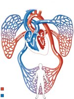
The heart is a muscular organ that functions as a double pump, circulating blood through two main circuits: the pulmonary circuit and the systemic circuit. It is essential for delivering oxygen and nutrients to tissues and removing waste products.
Pulmonary circuit: Right side of the heart pumps oxygen-poor blood to the lungs for gas exchange.
Systemic circuit: Left side of the heart pumps oxygen-rich blood to the rest of the body.
Heart Anatomy
External Structure and Orientation
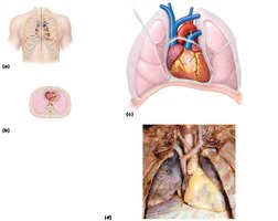
The heart is located in the mediastinum between the lungs, with its apex pointing to the left. It typically weighs 250–350 grams and is the largest organ of the mediastinum.
Apex: Inferior, pointed end directed leftward.
Base: Broad, superior surface facing posteriorly.
Gross Anatomy: External Features
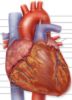
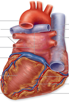
The heart has several external grooves (sulci) that mark the boundaries between its chambers:
Coronary sulcus: Encircles the heart, separating atria from ventricles.
Anterior and posterior interventricular sulci: Separate the right and left ventricles on the anterior and posterior surfaces, respectively.
Heart Chambers
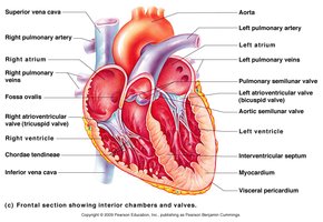
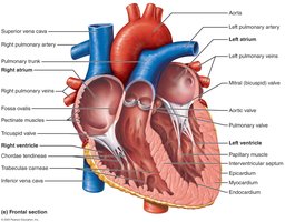
The heart consists of four chambers:
Right atrium: Receives oxygen-poor blood from the body via the superior and inferior venae cavae and the coronary sinus.
Right ventricle: Pumps blood into the pulmonary trunk toward the lungs.
Left atrium: Receives oxygen-rich blood from the lungs via the pulmonary veins.
Left ventricle: Pumps blood into the aorta for systemic circulation.
Internal Structures and Valves
Valves ensure unidirectional blood flow through the heart:
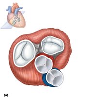
Atrioventricular (AV) valves: Tricuspid (right) and bicuspid/mitral (left) valves between atria and ventricles.
Semilunar valves: Pulmonary valve (right ventricle to pulmonary trunk) and aortic valve (left ventricle to aorta).
Heart Wall and Coverings
Pericardium
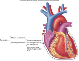
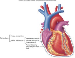
The pericardium is a double-walled sac that surrounds and protects the heart:
Fibrous pericardium: Tough, outer layer of dense connective tissue.
Serous pericardium: Thin, double-layered membrane (parietal and visceral layers) with a pericardial cavity filled with fluid to reduce friction.
Layers of the Heart Wall
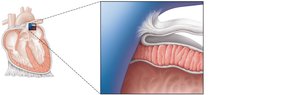
Epicardium: Visceral layer of the serous pericardium (outermost layer).
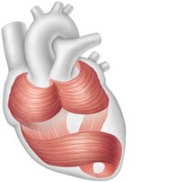
Myocardium: Thick, middle layer composed of cardiac muscle arranged in circular and spiral bundles.
Endocardium: Thin, inner lining of endothelium and connective tissue.
Microscopic Structure of Cardiac Muscle
Cardiac Muscle Tissue
Cardiac muscle tissue forms the myocardium and is responsible for the heart's contractile function.
Striated and contracts by the sliding filament mechanism.
Cells are short, branching, and typically have one or two nuclei.
Cells are joined by intercalated discs containing gap junctions and fasciae adherens for electrical and mechanical coupling.
Pathway of Blood Through the Heart
Sequential Flow
Blood flows through the heart in a specific sequence, ensuring separation of oxygen-poor and oxygen-rich blood:
Oxygen-poor blood enters the right atrium from the superior/inferior vena cava and coronary sinus.
Passes through the tricuspid valve into the right ventricle.
Pumped through the pulmonary semilunar valve into the pulmonary trunk and arteries to the lungs.
Oxygen-rich blood returns via pulmonary veins to the left atrium.
Passes through the mitral valve into the left ventricle.
Pumped through the aortic semilunar valve into the aorta and systemic circulation.
Heart Valves: Structure and Function
Valve Structure
Each valve consists of endocardium with a connective tissue core. The AV valves are supported by chordae tendineae and papillary muscles to prevent backflow during ventricular contraction.
Valve Function
AV valves open when atrial pressure exceeds ventricular pressure, allowing blood to flow into the ventricles.
During ventricular contraction, AV valves close to prevent regurgitation, and semilunar valves open to allow ejection of blood into arteries.
Conducting System of the Heart
Intrinsic Conduction System
The heart has an intrinsic ability to generate and conduct electrical impulses, coordinating rhythmic contractions:
Sinoatrial (SA) node: Pacemaker of the heart, initiates impulse.
Atrioventricular (AV) node: Delays impulse before passing to ventricles.
AV bundle (Bundle of His): Conducts impulse to bundle branches.
Bundle branches: Carry impulse through interventricular septum.
Purkinje fibers: Distribute impulse to ventricular myocardium.
Blood Supply to the Heart
Coronary Circulation
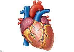
The heart muscle receives its blood supply from the right and left coronary arteries, which branch from the base of the aorta and run in the coronary sulcus. Major branches include:
Left coronary artery (LCA): Branches into the anterior interventricular artery (LAD) and circumflex artery.
Right coronary artery (RCA): Branches into the marginal artery and posterior interventricular artery (PDA).
Cardiac Veins
Deoxygenated blood from the heart wall is collected by cardiac veins and returned to the right atrium via the coronary sinus. Major veins include the great cardiac vein, middle cardiac vein, and small cardiac vein.
Heart Sounds
Valvular Closure
The "lub-dup" sounds of the heartbeat are produced by the closing of heart valves:
First sound (lub): Closure of AV valves.
Second sound (dup): Closure of semilunar valves.
Disorders of the Heart
Coronary Artery Disease
Atherosclerosis: Fatty deposits in coronary arteries.
Angina pectoris: Chest pain due to reduced blood flow.
Myocardial infarction: Heart attack caused by blocked coronary artery.
Heart Failure
Progressive weakening of the heart, leading to inadequate blood supply to tissues.
Congestive heart failure (CHF): Heart enlarges, pumping efficiency declines.
Disorders of the Conduction System
Arrhythmias: Abnormal heart rhythms.
Ventricular fibrillation: Rapid, uncoordinated contractions; can cause cardiac arrest.
Atrial fibrillation: Disorganized atrial impulses; increases risk of stroke.
Congenital Heart Defects
Most common defect: Ventricular septal defect (hole in the interventricular septum).
Other defects: Transposition of the great vessels, coarctation of the aorta, Tetralogy of Fallot.
Summary Table: Heart Chambers and Valves
Chamber | Receives Blood From | Pumps Blood To | Valve |
|---|---|---|---|
Right Atrium | Systemic veins (SVC, IVC, coronary sinus) | Right ventricle | Tricuspid (right AV) valve |
Right Ventricle | Right atrium | Pulmonary trunk | Pulmonary semilunar valve |
Left Atrium | Pulmonary veins | Left ventricle | Bicuspid (mitral, left AV) valve |
Left Ventricle | Left atrium | Aorta | Aortic semilunar valve |
