 Back
BackThe Cardiovascular System I: Blood & Vessels – Structure, Function, and Hemostasis
Study Guide - Smart Notes
 Tailored notes based on your materials, expanded with key definitions, examples, and context.
The Cardiovascular System: Overview
Major Components and Functions
The cardiovascular system is a closed network consisting of the heart, blood vessels, and blood. Its primary function is to transport nutrients, gases, hormones, and waste products throughout the body, ensuring cellular homeostasis and communication.
Heart: The muscular pump that generates pressure to move blood through vessels.
Blood Vessels: Tubular structures (arteries, veins, capillaries) that direct blood flow.
Blood: The transport medium carrying cells, nutrients, gases, and waste.
General functions include delivery of oxygen and nutrients, removal of metabolic wastes, hormone transport, immune defense, and regulation of body temperature and pH.
Blood: Composition and Properties
Physical and Chemical Characteristics
State: Liquid, slightly more viscous than water.
Color: Bright red when oxygenated, purplish when deoxygenated (due to hemoglobin).
pH: Slightly alkaline, averaging 7.4 (range: 7.35–7.45). Venous blood is more acidic than arterial blood.
Volume: 6–8% of body weight (4–6 quarts in adults).
Temperature: ~37°C.
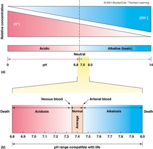
Clinical Note: Blood pH below 7.35 is termed acidosis; above 7.45 is alkalosis. Life is not compatible with pH below 6.8 or above 8.0.
Blood Constituents
Plasma: Straw-colored liquid (55% of blood), mostly water (91.5%), containing proteins, nutrients, electrolytes, gases, and waste products.
Formed Elements: Cellular portion (45%): erythrocytes (RBCs), leukocytes (WBCs), and platelets (thrombocytes).
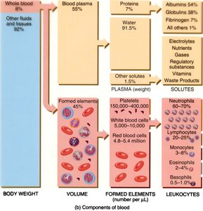
Plasma Proteins
Albumins: Maintain osmotic pressure, transport substances.
Globulins: Transport, immune functions (antibodies).
Fibrinogen: Precursor to fibrin in blood clotting.
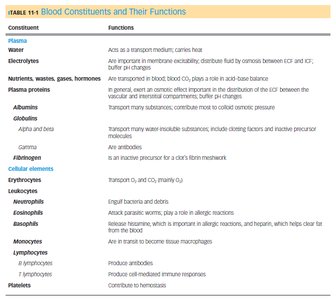
Formed Elements of Blood
Erythrocytes (Red Blood Cells)
Erythrocytes are biconcave, anucleate cells specialized for gas transport. Their shape increases surface area and flexibility, allowing passage through capillaries. Each cell contains over 250 million hemoglobin molecules, which bind oxygen and carbon dioxide.
Hemoglobin: Composed of four polypeptide subunits, each with an iron-containing heme group. Each hemoglobin can bind four O2 molecules.
Hematocrit: Percentage of blood volume occupied by RBCs (males: 40–54%, females: 37–47%).
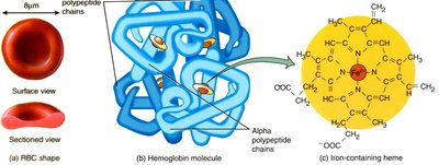
Leukocytes (White Blood Cells)
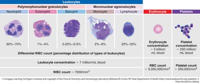
Leukocytes are involved in immune defense and tissue maintenance. They are classified as granulocytes (neutrophils, eosinophils, basophils) and agranulocytes (lymphocytes, monocytes).
Neutrophils: Phagocytize bacteria and debris.
Eosinophils: Combat parasites, modulate allergic responses.
Basophils: Release histamine and heparin in inflammation/allergy.
Lymphocytes: B cells (antibody production), T cells (cell-mediated immunity).
Monocytes: Differentiate into macrophages for phagocytosis.
Platelets (Thrombocytes)
Platelets are cell fragments essential for hemostasis (blood clotting). They aggregate at injury sites, forming a temporary plug and releasing factors that promote coagulation.
Hematopoiesis and Erythropoiesis
Blood Cell Formation
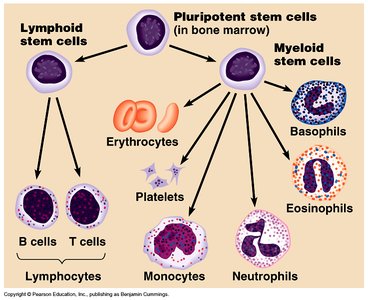
All blood cells originate from pluripotent hematopoietic stem cells in the bone marrow. Differentiation follows two main lineages: myeloid (producing erythrocytes, platelets, granulocytes, monocytes) and lymphoid (producing lymphocytes).
Erythropoiesis
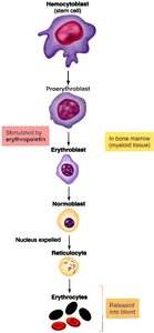
Erythropoiesis is the process of RBC production, regulated primarily by erythropoietin (EPO), a hormone released in response to hypoxia. The maturation sequence includes proerythroblast, erythroblast, normoblast (nucleus expelled), reticulocyte, and mature erythrocyte.
Blood Cell Recycling and Jaundice
Erythrocyte Recycling
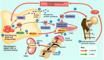
After ~120 days, aged RBCs are phagocytosed by macrophages in the spleen and liver. Hemoglobin is broken down: globin to amino acids, heme to iron (recycled) and bilirubin (excreted in bile).
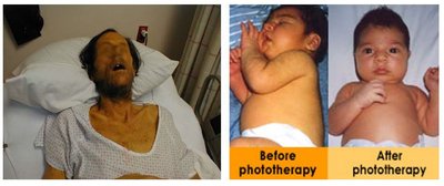
Jaundice
Excess bilirubin (hyperbilirubinemia) causes jaundice, characterized by yellowing of the skin and eyes. It may result from liver dysfunction or rapid RBC breakdown.
Blood Vessels: Structure and Function
General Structure of Blood Vessels
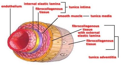
Except for capillaries, blood vessels have three layers (tunics):
Tunica intima: Endothelium and internal elastic lamina (innermost layer).
Tunica media: Smooth muscle and elastic fibers (middle layer; thickest in arteries).
Tunica adventitia: Connective tissue (outermost layer; thickest in veins).
Arteries vs. Veins
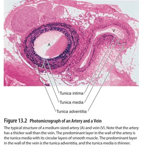
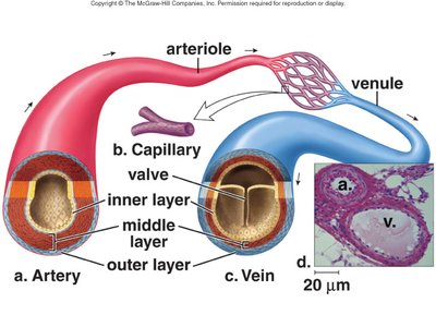
Arteries: Thick tunica media, high elasticity, function as resistance vessels (maintain blood pressure).
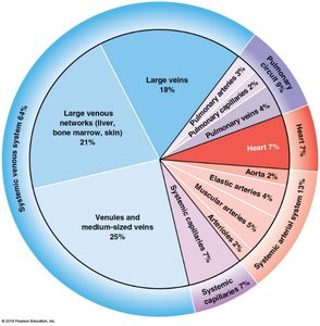
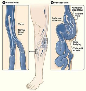
Veins: Thinner walls, larger lumen, function as compliance vessels (blood reservoir), contain valves to prevent backflow.
Capillaries: Types and Functions
Capillary Structure and Exchange
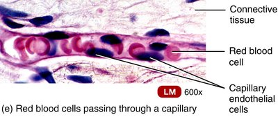
Capillaries are the smallest blood vessels, consisting of a single layer of endothelium. They facilitate exchange of gases, nutrients, and wastes between blood and tissues.
Types of Capillaries
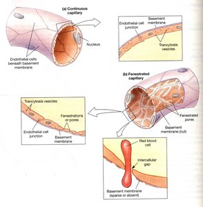
Continuous: Most common; uninterrupted endothelium. Found in muscle, nervous tissue.
Fenestrated: Have pores for rapid exchange; found in kidneys, small intestine.
Sinusoidal (discontinuous): Large gaps; allow passage of cells and proteins. Found in liver, spleen, bone marrow.
Venous Return and Blood Distribution
Mechanisms of Venous Return
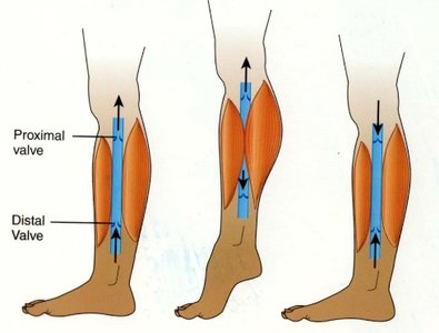
Muscular Compression: Skeletal muscle contractions squeeze veins, propelling blood toward the heart (aided by venous valves).
Respiratory Pump: Changes in thoracic pressure during breathing enhance venous return.
Venous Valves: Prevent backflow, especially in limbs.
Hemostasis: Prevention of Blood Loss
Hemostasis Overview
Hemostasis is the process that stops bleeding after vessel injury. It involves three main steps:
Vascular Spasm: Immediate vasoconstriction reduces blood flow.
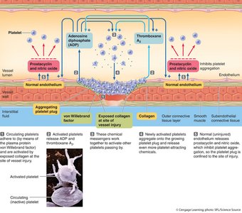
Platelet Plug Formation: Platelets adhere to exposed collagen, aggregate, and release chemicals (ADP, thromboxane A2) to recruit more platelets.
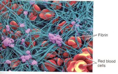
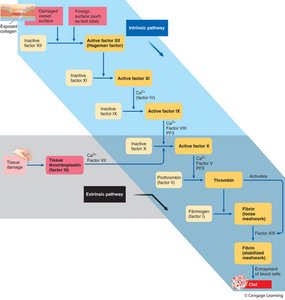
Coagulation (Clotting): Cascade of enzymatic reactions converts fibrinogen to fibrin, forming a stable clot.
Clot Retraction and Removal
After vessel repair, the clot is dissolved by plasmin (fibrinolysis), and phagocytes remove debris. Platelets contract to shrink the clot and draw wound edges together.
Summary Table: Blood Constituents and Functions
Constituent | Function |
|---|---|
Plasma | Transport medium, carries heat, maintains osmotic balance |
Albumins | Osmotic pressure, transport substances |
Globulins | Immune functions, transport |
Fibrinogen | Clot formation |
Erythrocytes | Transport O2 and CO2 |
Leukocytes | Immune defense |
Platelets | Hemostasis (clotting) |
Key Equations
Hematocrit:
Osmotic Pressure (van't Hoff's Law):
Additional info: This guide integrates foundational concepts from human anatomy and physiology, focusing on blood and vessels, and is suitable for ANP college-level study.
