Back
BackThe Cardiovascular System II: The Blood Vessels – Study Notes
Study Guide - Smart Notes
 Tailored notes based on your materials, expanded with key definitions, examples, and context.
Introduction to Vasculature (Blood Vessels)
General Functions of Blood Vessels
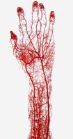
The vasculature, or network of blood vessels, is essential for transporting blood throughout the body, regulating blood flow to tissues, and controlling blood pressure. Blood vessels form a closed circuit that ensures efficient delivery of oxygen, nutrients, and removal of waste products.
Transport blood to and from the heart and tissues.
Regulate blood flow by constricting or dilating.
Control blood pressure through vessel diameter and elasticity.
Types of Blood Vessels
Arteries: Distribution System
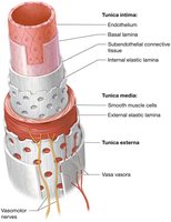
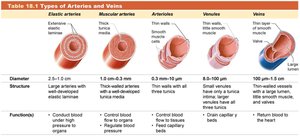
Arteries carry blood away from the heart and are classified based on their structure and function. Their walls are composed of three layers, each with distinct properties:
Tunica intima: Innermost layer; smooth endothelium for reduced friction, supported by connective tissue with elastic and collagen fibers.
Tunica media: Middle layer; contains smooth muscle and elastic connective tissue, allowing for vasoconstriction and vasodilation.
Tunica externa: Outermost layer; thin connective tissue that anchors the vessel to surrounding structures.
Classification of Arteries
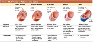
Elastic arteries: Largest diameter, closest to the heart, withstand highest pressure (e.g., aorta).
Muscular arteries: Intermediate diameter, well-developed tunica media, supply organs.
Arterioles: Smallest arteries, thin walls, regulate blood flow to tissues; have branches called metarterioles that feed capillary beds.
Type | Diameter | Structure | Function |
|---|---|---|---|
Elastic arteries | 2.5–1 cm | Large elastic lamina, thick tunica media | Conduct blood under high pressure |
Muscular arteries | 1 cm–0.3 mm | Thick-walled, well-developed tunica media | Regulate blood flow to organs |
Arterioles | 0.3 mm–10 μm | Thin walls, less muscle | Control blood flow to capillary beds |
Venules | 8–100 μm | Thin walls, little muscle | Drain capillary beds |
Veins | 100 μm–1.5 cm | Thin-walled, valves present | Return blood to the heart |
Capillaries: Exchange System
Capillaries are the smallest blood vessels, forming networks that connect arterioles and venules. Their thin walls (simple squamous epithelium) facilitate the exchange of gases, nutrients, and wastes between blood and tissues. Flow is regulated by precapillary sphincters, which open when cells are low in oxygen and nutrients.
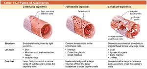
Continuous capillaries: Least permeable, permit a narrow range of substances to cross (e.g., muscle, skin, brain).
Fenestrated capillaries: Moderately permeable, allow larger volumes of fluid and substances (e.g., kidneys, endocrine glands).
Sinusoidal capillaries: Most permeable, allow large substances such as cells to cross (e.g., liver, spleen).
Type | Structure | Location | Function |
|---|---|---|---|
Continuous | Tight junctions | Muscle, skin, brain | Least leaky, selective exchange |
Fenestrated | Fenestrations (pores) | Kidneys, endocrine glands | Moderately leaky, larger exchange |
Sinusoidal | Large gaps | Liver, spleen | Leakiest, allow cells to cross |
Veins: Collection System
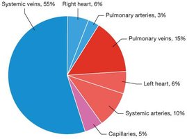
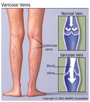
Veins return blood from capillary beds to the heart. Their walls are similar to arteries but thinner, with less muscle and elastic tissue. Many veins contain venous valves to prevent backflow. Veins act as blood reservoirs, containing about 70% of the body's blood volume, which helps maintain blood pressure during blood loss.
Venules: Smallest veins, drain blood from capillary beds, thin walls.
Veins: Larger vessels, thin walls, valves present, return blood to the heart.
Blood Vessel Disorders
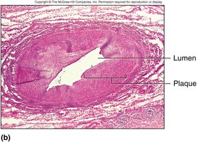
Atherosclerosis: Accumulation of fatty material (plaque) inside an artery, narrowing the lumen and restricting blood flow.
Arteriosclerosis: Hardening of arteries, often a later stage of atherosclerosis.
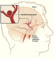
Aneurysm: Dilation and weakening of an artery wall, forming a pulsating sac that may rupture.
Phlebitis: Inflammation of a vein.
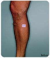
Varicose veins: Abnormal dilation of superficial veins, often in the legs; risk factors include heredity, hormones, obesity, aging, and prolonged standing.
Physiology of Blood Flow (Hemodynamics)
Main Factors Influencing Blood Pressure
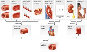
Hemodynamics refers to the study of blood flow and the forces involved. Blood pressure is influenced by three main factors:
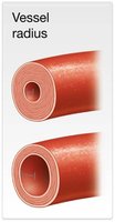
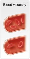
Peripheral resistance: Resistance to blood flow, affected by vessel location, radius, viscosity, and length. Resistance increases with distance from the heart, smaller vessel radius, higher viscosity, and longer vessels.
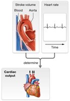
Cardiac output (CO): The volume of blood pumped by the heart per minute. Increased CO raises blood pressure.
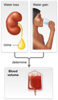
Blood volume: Directly related to the amount of water in the blood; increased volume raises blood pressure.
Blood Pressure
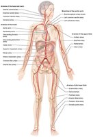
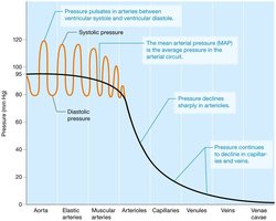
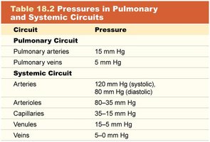
Blood pressure is the force exerted by blood against vessel walls, typically measured in the brachial artery. It is expressed as systolic/diastolic pressure (e.g., 120/80 mm Hg). Pulse pressure is the difference between systolic and diastolic pressures. Blood pressure decreases as blood moves through the arterial system and is nearly zero in veins.
Systolic pressure: 110–120 mm Hg
Diastolic pressure: 70–80 mm Hg
Pulse pressure: ~40 mm Hg
Mean Arterial Pressure (MAP): Average pressure in systemic arteries during the cardiac cycle.
MAP is calculated as:
(Average MAP ≈ 93 mm Hg)
Maintenance of Blood Pressure
Nervous system: Sympathetic and parasympathetic controls, including the baroreceptor reflex (mechanoreceptors in vessels).
Urinary system: Kidneys regulate blood volume via urine formation.
Endocrine system: Hormones (e.g., antidiuretic hormone, aldosterone) control cardiac output, vessel diameter, and kidney function.
Disorders of Blood Pressure
Hypertension: High blood pressure, often asymptomatic but increases risk for coronary artery disease, stroke, heart failure, dementia, kidney disease, and vascular disease.
Hypotension: Low blood pressure (systolic < 90 mm Hg, diastolic < 60 mm Hg); can cause dizziness, fainting, or organ failure in severe cases.
Capillary Exchange (Microcirculation)
Mechanisms of Capillary Exchange
Capillary exchange allows the transfer of water, electrolytes, gases, wastes, glucose, and lipids between blood and tissues. This process is governed by hydrostatic and osmotic pressures:
Hydrostatic Pressure (HP): The force exerted by blood against capillary walls, pushing fluid out.
Osmotic Pressure (OP): The movement of water toward higher solute concentration, regulated by plasma proteins (e.g., albumin).
Processes Involved
Diffusion: Movement of substances down their concentration gradients; facilitated by fenestrations, lipid solubility, and transcytosis. Oxygen and nutrients leave blood at the arteriole end; wastes and CO2 enter at the venule end.
Filtration: Movement of fluid out of capillaries at the arteriole end (HP > OP); reabsorption at the venule end (OP > HP).
Osmosis: Water moves toward higher solute concentration.
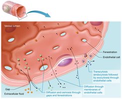
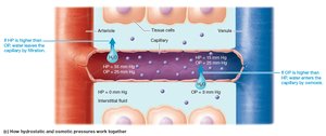
Normally, more fluid leaves capillaries than returns; excess is collected by lymphatic capillaries and returned to circulation.
Edema
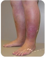
Edema is swelling caused by excess fluid in tissues, often due to decreased filtration at venules or heart failure (right ventricle cannot pump blood efficiently).
Anatomy of Systemic Arteries and Veins
Hepatic Portal System
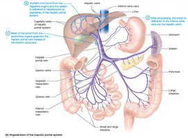
The hepatic portal system allows the liver to monitor and process nutrients and chemicals absorbed from the gastrointestinal tract. Blood from digestive organs is collected into the hepatic portal vein, which carries nutrient-rich (but oxygen-poor) blood to the liver. The liver stores nutrients, detoxifies substances, and destroys bacteria before blood returns to the systemic circulation via the hepatic veins and inferior vena cava.
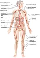
Celiac artery: Supplies stomach, liver, duodenum, pancreas, spleen.
Superior mesenteric artery: Supplies pancreas, small and large intestines.
Portal system begins and ends in capillaries.
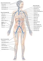
Hepatic portal vein carries blood to the liver, where it breaks into capillary networks (sinusoids).
Liver receives oxygenated blood from the hepatic artery.