 Back
BackThe Cardiovascular System: Structure and Function of the Heart
Study Guide - Smart Notes
 Tailored notes based on your materials, expanded with key definitions, examples, and context.
The Cardiovascular System
Introduction
The cardiovascular system is essential for transporting blood, nutrients, gases, and wastes throughout the body. It consists of the heart, which acts as a pump, and an extensive network of blood vessels. The study of the heart and its diseases is known as cardiology.
Heart: Pumps blood through approximately 60,000 miles of blood vessels.
Blood vessels: Include arteries, veins, and capillaries.
Cardiology: The study of the heart and its disorders.
Overview of Cardiovascular Components
Blood Vessels
Arteries: Carry blood away from the heart (most carry oxygenated blood).
Veins: Carry blood toward the heart (most carry deoxygenated blood).
Capillaries: Sites of exchange between blood and tissues or air in the lungs.
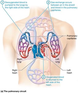
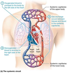
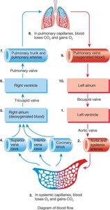
Pulmonary and Systemic Circulation
The heart supports two main circulatory circuits:
Pulmonary circulation: Carries deoxygenated blood from the right side of the heart to the lungs for gas exchange, then returns oxygenated blood to the left side of the heart.
Systemic circulation: Delivers oxygenated blood from the left side of the heart to the body, where it exchanges gases, nutrients, and wastes, then returns deoxygenated blood to the right side of the heart.
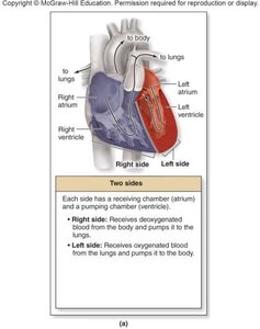
The Heart’s Four Chambers
The heart is divided into four chambers:
Right atrium and left atrium: Superior chambers that receive blood.
Right ventricle and left ventricle: Inferior chambers that pump blood away from the heart.
The left side handles oxygenated blood; the right side handles deoxygenated blood.
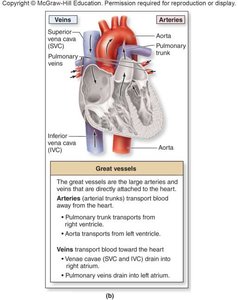
Great Vessels of the Heart
Pulmonary trunk: Transports deoxygenated blood from the right ventricle to the lungs.
Aorta: Transports oxygenated blood from the left ventricle to the body.
Superior and inferior vena cava: Return deoxygenated blood from the body to the right atrium.
Pulmonary veins: Return oxygenated blood from the lungs to the left atrium.
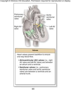
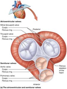
Heart Valves
The heart contains two sets of valves to ensure one-way blood flow:
Atrioventricular (AV) valves: Between atria and ventricles (right AV = tricuspid, left AV = bicuspid/mitral).
Semilunar (SL) valves: Between ventricles and arterial trunks (pulmonary and aortic semilunar valves).
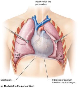
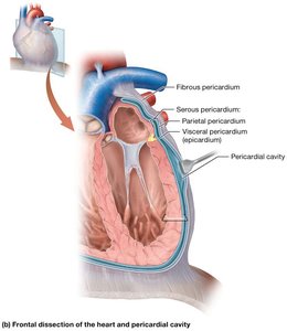
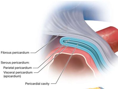
Pericardium and Heart Wall Structure
Pericardium
The pericardium is a double-walled sac that encloses the heart and anchors it in place.
Fibrous pericardium: Dense connective tissue that protects and anchors the heart.
Serous pericardium: Thin membrane with two layers (parietal and visceral/epicardium) and a pericardial cavity filled with fluid to reduce friction.
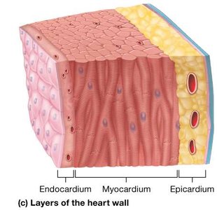
Layers of the Heart Wall
Epicardium: Outer layer (visceral pericardium).
Myocardium: Middle, muscular layer responsible for contraction.
Endocardium: Inner layer lining the chambers and valves.
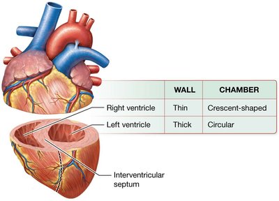
Thickness of the Heart Wall
Atria have thin walls; ventricles have thicker walls due to higher pressure requirements.
The left ventricle has the thickest wall to pump blood throughout the body.
Heart Valve Function
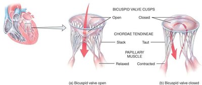
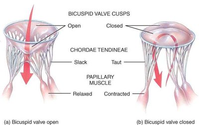
Atrioventricular Valves
Open when ventricular pressure is lower than atrial pressure (ventricles relaxed).
Close when ventricles contract, preventing backflow into the atria.
Chordae tendineae and papillary muscles prevent valve prolapse.
Semilunar Valves
Open during ventricular contraction to allow blood flow into the pulmonary trunk or aorta.
Close during ventricular relaxation to prevent backflow into the ventricles.
Heart Valve Disorders
Stenosis: Narrowing of a valve, restricting blood flow.
Insufficiency (incompetence): Failure of a valve to close completely, causing backflow.
Heart murmurs: Abnormal sounds due to turbulent blood flow through defective valves.
Blood and Coronary Circulation
Blood Flow Pathway
Deoxygenated blood enters the right atrium, passes to the right ventricle, and is pumped to the lungs via the pulmonary trunk.
Oxygenated blood returns from the lungs to the left atrium, passes to the left ventricle, and is pumped to the body via the aorta.
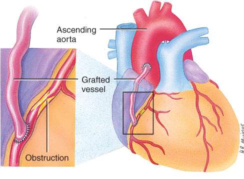
Coronary Circulation
Supplies blood to the heart muscle itself.
Coronary arteries branch from the aorta; many anastomoses provide alternate routes for blood flow.
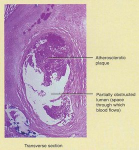
Coronary Artery Disease (CAD)
Caused by narrowing of coronary arteries due to atherosclerosis, spasm, or clot.
Treatments include drugs, coronary artery bypass graft (CABG), angioplasty, and stenting.
Histology and Physiology of Cardiac Muscle
Cardiac Muscle Structure
Short, branched fibers connected by intercalated discs (desmosomes and gap junctions).
Orderly arrangement of actin and myosin in sarcomeres.
Requires extracellular calcium for contraction.
Types of Cardiac Muscle Cells
Contractile cells: Responsible for contraction and relaxation of the heart.
Autorhythmic cells: Generate and conduct electrical impulses (pacemaker and conduction system).
Properties of Cardiac Muscle
Self-excitable (can generate its own action potentials).
Conducts impulses throughout the heart.
Contracts and relaxes rhythmically.
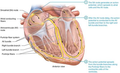
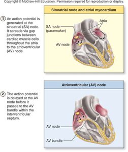
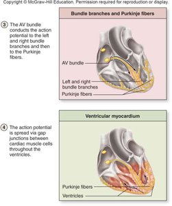
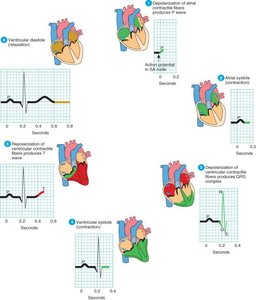
Conduction System of the Heart
Components and Function
The conduction system coordinates the contraction of the heart muscle, ensuring efficient pumping.
Sinoatrial (SA) node: Pacemaker, initiates impulses (90–120/min).
Atrioventricular (AV) node: Delays impulse, allows atrial contraction (40–60/min).
AV bundle (Bundle of His) and Purkinje fibers: Conduct impulses to ventricles (20–40/min).
Heart Rate and Its Regulation
Normal Heart Rate
Normal range: 60–90 beats/min; average: 70–75 beats/min.
Tachycardia: HR > 100 beats/min.
Bradycardia: HR < 60 beats/min.
Factors Affecting Heart Rate
Nervous system: Sympathetic nerves increase HR and force; parasympathetic (vagus) decreases HR and force.
Temperature: Increased temperature raises HR; decreased temperature lowers HR.
Respiration: HR increases during inspiration, decreases during expiration (sinus arrhythmia).
Hormones: Epinephrine, norepinephrine, thyroid hormones increase HR; acetylcholine decreases HR.
Drugs: Digitalis, ouabain, quinidine, dopamine affect HR.
Emotional states: Fear, pain, anxiety, anger, excitement increase HR; sudden grief decreases HR.
Exercise and fever: Both increase HR.
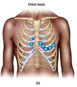
Electrocardiogram (ECG)
Principles and Recording
ECG records the electrical activity of the heart using electrodes placed on the skin.
Standard limb leads (I, II, III), augmented limb leads (aVR, aVL, aVF), and precordial (chest) leads (V1–V6) are used.
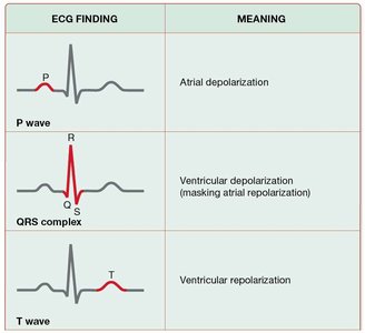
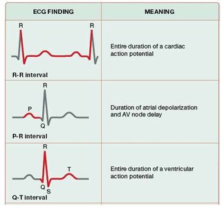
ECG Waves and Intervals
P wave: Atrial depolarization.
PR interval: Time for impulse conduction from atria to ventricles.
QRS complex: Ventricular depolarization.
T wave: Ventricular repolarization.
Uses of ECG
Assess heart rate, rhythm, conduction abnormalities, heart size, effects of ionic changes and drugs, myocardial ischemia or infarction, and other heart diseases.
ECG Abnormalities
Bradycardia: Slow heart rate.
Tachycardia: Fast heart rate.
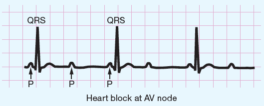
Conduction block: Delay or block in impulse conduction, often at the AV node.
Arrhythmia: Irregular heart rhythm, including atrial and ventricular types.
Summary Table: Heart Valves
Valve | Location | Function |
|---|---|---|
Tricuspid (Right AV) | Between right atrium and right ventricle | Prevents backflow into right atrium |
Bicuspid/Mitral (Left AV) | Between left atrium and left ventricle | Prevents backflow into left atrium |
Pulmonary Semilunar | Between right ventricle and pulmonary trunk | Prevents backflow into right ventricle |
Aortic Semilunar | Between left ventricle and aorta | Prevents backflow into left ventricle |
Summary Table: ECG Waves and Intervals
ECG Finding | Meaning |
|---|---|
P wave | Atrial depolarization |
QRS complex | Ventricular depolarization |
T wave | Ventricular repolarization |
PR interval | Conduction time from atria to ventricles |
QT interval | Duration of ventricular action potential |
