 Back
BackThe Cardiovascular System: Structure, Function, and Circulation
Study Guide - Smart Notes
 Tailored notes based on your materials, expanded with key definitions, examples, and context.
The Cardiovascular System
Heart Structure and Location
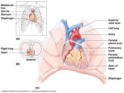
The heart is a muscular organ about the size of your fist, located in the mediastinum (the space between the lungs, backbone, and sternum). It is central to the circulatory system, pumping blood throughout the body. - Size: Approximately 9x14 cm. - Location: Mediastinum, between the lungs. - Function: Pumps blood to supply oxygen and nutrients to tissues and remove waste products. 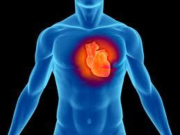
Heart Coverings: The Pericardium
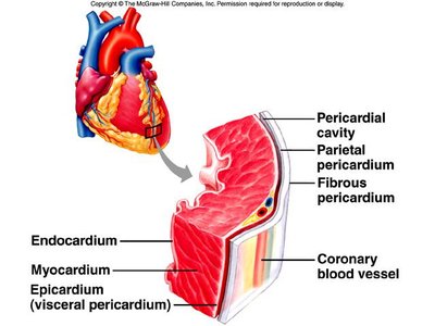
The heart is enclosed by the pericardium, a double-walled sac that protects and anchors the heart, preventing overfilling and reducing friction during contractions. - Parietal layer: Outermost covering. - Visceral layer: Lines the surface of the heart. - Pericardial fluid: Lubricates and protects the heart. 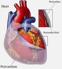
Parts and Chambers of the Heart
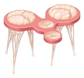
The heart consists of four chambers and several key structures that ensure efficient blood flow. - Atria: Receiving chambers where blood enters the heart. - Ventricles: Pumping chambers where blood leaves the heart. - Valves: Prevent backflow and ensure one-way blood flow. - Chordae tendineae: Cord-like tendons anchoring the AV valves, resisting blood pressure during ventricular contraction. - Septum: Myocardial wall separating right and left ventricles. 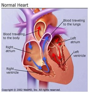
Heart Valves
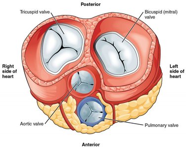
Valves ensure blood flows in one direction only, preventing backflow between contractions. - Atrioventricular (AV) valves: Between atrium and ventricle (Tricuspid on right, Bicuspid/Mitral on left). - Semilunar valves: Between ventricle and outbound artery (Pulmonary on right, Aortic on left). - Function: AV valves close during ventricular contraction; semilunar valves close during ventricular relaxation. 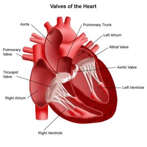
Blood Vessels Attached to the Heart
Several major blood vessels are directly connected to the heart, facilitating circulation. - Aorta: Largest artery, carries oxygenated blood from heart to body. - Pulmonary veins: Carry oxygenated blood from lungs to heart. - Pulmonary artery: Carries deoxygenated blood from heart to lungs. - Vena cavas: Veins carrying deoxygenated blood from body to heart.
Heart Circuits
Pulmonary and Systemic Circulation
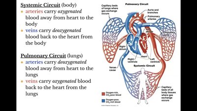
The heart acts as two pumps: one for the lungs (pulmonary circulation) and one for the body (systemic circulation). - Pulmonary circuit: Right ventricle → pulmonary arteries → lung capillaries → pulmonary veins → left atrium. - Systemic circuit: Left ventricle → arteries → body capillaries → veins → right atrium. 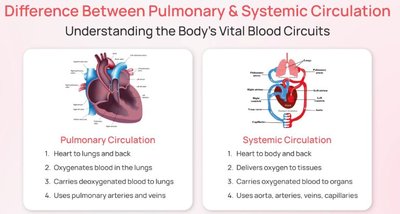
Blood Vessels
Types of Blood Vessels
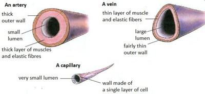
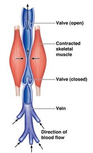
Blood vessels are classified into arteries, veins, and capillaries, each with distinct structure and function. - Arteries: Strong, elastic vessels carrying blood away from the heart; thick walls of smooth muscle and elastic fibers; smaller lumen. - Veins: Thinner, less muscular vessels carrying blood toward the heart; larger lumen; contain valves to prevent backflow. - Capillaries: Smallest vessels; walls made of a single layer of squamous cells; site of gas and nutrient exchange. 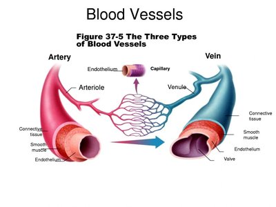

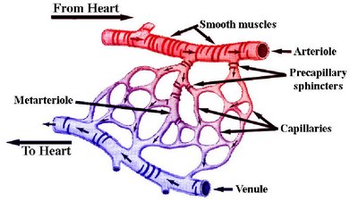
Capillary Function
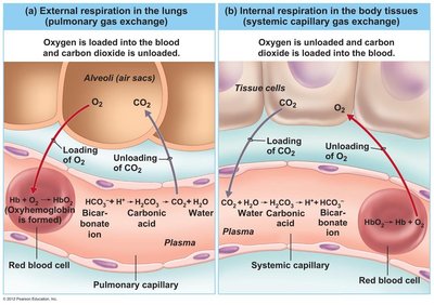
Capillaries penetrate nearly all tissues, allowing exchange of gases (O2 and CO2) and nutrients between blood and tissues. - Wall composition: Single layer of squamous cells for rapid diffusion. - Critical function: Exchange of materials between blood and tissues. 
Cardiac Conduction System
Electrical Impulse Generation
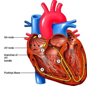
The cardiac conduction system (nodal system) is a group of specialized cardiac muscle cells that send signals to the heart muscle, causing it to contract. - Components: SA node, AV node, bundle of His, bundle branches, Purkinje fibers. - Function: Initiates and coordinates heart contractions independently of the nervous system. 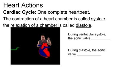
Regulation of Cardiac Cycle
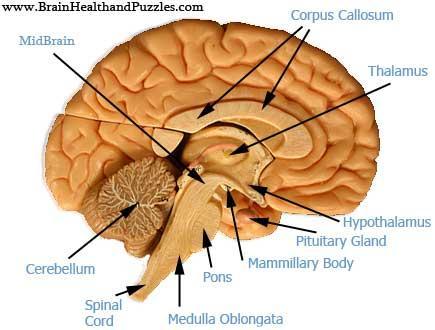
Heart rate is controlled by the cardiac center within the medulla oblongata, adjusting in response to changes in the body's state (homeostasis). 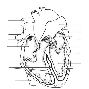
Electrocardiograms (ECG)
Measuring Heart Activity
Electrocardiograms (ECGs) measure electrical impulses from the heart, creating a graph of the output. - P wave: Atria contract. - QRS wave: Ventricles contract. - T wave: Ventricles relax and reset.
Heart Rate
- Normal resting heart rate: 60-100 beats per minute. - Bradycardia: Heart rate below 60 bpm. - Tachycardia: Heart rate above 100 bpm.
Coronary Circulation
Supplying the Heart Muscle
Coronary circulation is the specialized vascular network supplying oxygenated blood to the myocardium (heart muscle). - Coronary arteries: Supply oxygenated blood to heart muscle. - Coronary veins: Carry away deoxygenated blood.
Heart Sounds
Normal Heart Sounds
- S1 (Lub): Closure of atrioventricular (mitral and tricuspid) valves at the beginning of systole. - S2 (Dub): Closure of semilunar (aortic and pulmonic) valves at the beginning of diastole.
Blood Pressure
Definition and Measurement
Blood pressure is the force of blood pushing against the walls of arteries as the heart pumps it throughout the body. - Systolic pressure: Peak pressure during ventricular contraction. - Diastolic pressure: Low pressure during ventricular refilling. - Normal range: 120-140 / 80-90 mmHg.
Cardiovascular Disorders
Atherosclerosis
Atherosclerosis is a buildup of lipids, calcium, or cell debris that restricts blood flow, causing higher blood pressure and potentially leading to heart attacks if a blockage occurs in a coronary artery.
Aneurysm
An aneurysm is a weakening of a blood vessel wall, causing it to bulge and potentially burst or form a thrombus. Risk factors include high blood pressure, atherosclerosis, and smoking.
Heart Attack
A heart attack is caused by a blockage in a coronary artery, disrupting the flow of oxygenated blood to the heart muscle.
Coronary Bypass and Angioplasty
- Coronary bypass: Transplants a vein to reroute blood around a blockage. - Angioplasty: Balloon inserted and inflated to expand a blocked vessel; a stent may be used to keep it open.
Hypertrophic Cardiomyopathy
Abnormal thickening of the heart muscle wall, decreasing the heart’s ability to pump blood. Usually inherited or occurs with age.
Hemostasis and Blood Clotting
Stages of Hemostasis
Hemostasis is the stoppage of bleeding from a break in a blood vessel. - Stage 1: Platelets attach to the vessel wall, forming a platelet plug. - Stage 2: Platelets activate thrombin, which activates fibrin strands that stick to the plug. - Stage 3: Fibrin proteins cause red blood cells to stick to the clot, sealing the vessel until it heals.
Blood Clotting Disorders
- Hemophilia: Hereditary disorder impairing blood clotting; platelet plug forms but fibrin does not. - Thrombus: Blood clot in an unbroken vessel. - Embolus: Thrombus that breaks away and clogs a vessel elsewhere (e.g., stroke). - Hematoma: Swelling of clotted blood within tissue; bruise colors result from hemoglobin breakdown.
Fetal Circulation
Unique Features of Fetal Circulation
In a fetus, the lungs are nonfunctional and are bypassed by the foramen ovale, a hole allowing blood to pass directly from the right atrium to the left atrium. The foramen ovale closes at birth. - Umbilical cord: Contains one vein (oxygenated blood to fetus) and two arteries (deoxygenated blood from fetus to mother). - Fetal hemoglobin: Greater affinity for oxygen than maternal hemoglobin.
Summary Table: Heart Valves and Circuits
Valve | Location | Circuit | Function |
|---|---|---|---|
Tricuspid (AV) | Right atrium & ventricle | Pulmonary | Prevents backflow to atrium |
Bicuspid/Mitral (AV) | Left atrium & ventricle | Systemic | Prevents backflow to atrium |
Pulmonary Semilunar | Right ventricle & pulmonary artery | Pulmonary | Prevents backflow to ventricle |
Aortic Semilunar | Left ventricle & aorta | Systemic | Prevents backflow to ventricle |
Key Equations
Blood Pressure
Cardiac Output
Summary Table: Blood Vessel Structure
Type | Lumen Size | Wall Structure | Function |
|---|---|---|---|
Artery | Small | Thick muscle & elastic fibers | Carry blood away from heart |
Vein | Large | Thin muscle & elastic fibers, valves | Carry blood toward heart |
Capillary | Very small | Single layer of squamous cells | Exchange of gases & nutrients |
