 Back
BackThe Cardiovascular System: The Heart and Blood Vessels
Study Guide - Smart Notes
 Tailored notes based on your materials, expanded with key definitions, examples, and context.
The Cardiovascular System
Overview of the Cardiovascular System
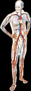
The cardiovascular system is responsible for the transport of blood, nutrients, gases, and wastes throughout the body. It consists of the heart, blood vessels, and blood. The heart acts as a pump to propel blood through a closed system of vessels, ensuring efficient circulation to all tissues.
Chapter 18: The Heart
Anatomical Location of the Heart
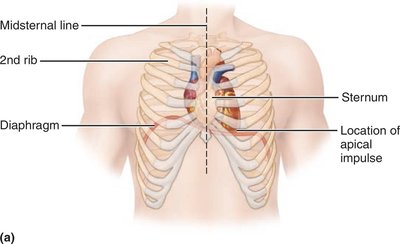
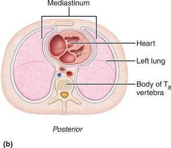
The heart is located in the mediastinum, the central compartment of the thoracic cavity, between the sternum and vertebral column, and between the lungs. It rests on the diaphragm and is bordered superiorly by the first rib.
Cardiopulmonary Resuscitation (CPR)
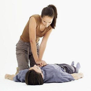
CPR is a life-saving technique that combines chest compressions with artificial ventilation to maintain circulatory flow and oxygenation during cardiac arrest. The heart's position between the sternum and vertebral column allows effective compressions to propel blood.
Layers of the Heart Wall
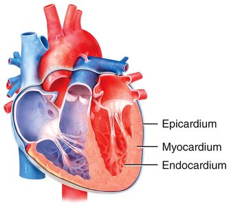
The heart wall consists of three layers:
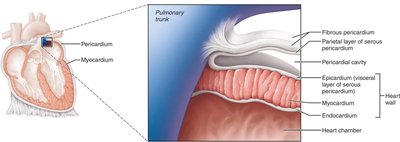
Epicardium: The outermost layer, also known as the visceral layer of the serous pericardium.
Myocardium: The thick, muscular middle layer responsible for contraction and pumping action.
Endocardium: The innermost layer, lining the heart chambers and covering the valves.
Pericardium and Pericardial Cavity
The heart is enclosed in a double-walled sac called the pericardium, which consists of:
Fibrous pericardium: Tough, dense connective tissue that protects and anchors the heart.
Serous pericardium: A thin, double-layered membrane (parietal and visceral layers) with a pericardial cavity filled with fluid to reduce friction during heartbeats.
Gross Anatomy of the Heart
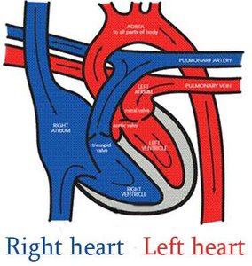
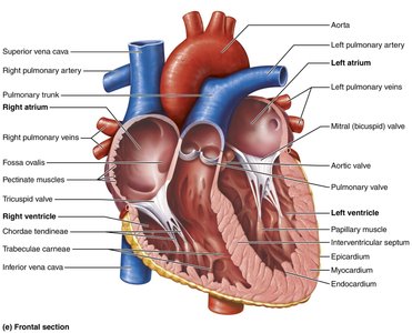
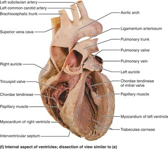
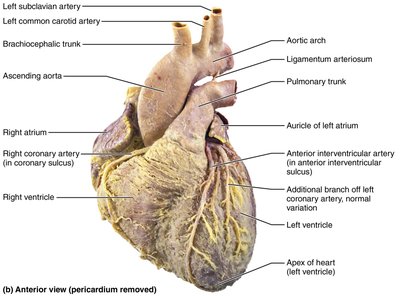
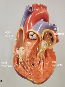
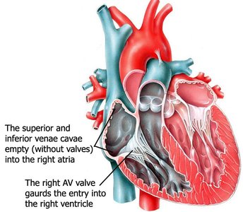
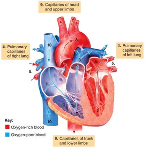
The heart is divided into four chambers: two atria (upper chambers) and two ventricles (lower chambers). The right side receives deoxygenated blood and pumps it to the lungs (pulmonary circulation), while the left side receives oxygenated blood and pumps it to the body (systemic circulation).
Comparison of Heart Chambers
The atria are thin-walled chambers that receive blood, while the ventricles are thick-walled and serve as the main pumps. The left ventricle has the thickest myocardium because it must generate high pressure to pump blood throughout the systemic circuit.
Heart Valves
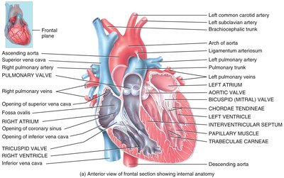
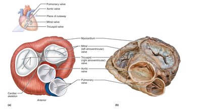
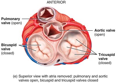
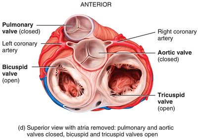
Valves ensure unidirectional blood flow through the heart:
Atrioventricular (AV) valves: Tricuspid (right) and bicuspid/mitral (left) valves between atria and ventricles.
Semilunar (SL) valves: Pulmonary and aortic valves at the exits of the ventricles.
Blood Flow Through the Heart
Blood flows through the heart in a specific sequence, passing through the right atrium, right ventricle, pulmonary arteries, lungs, pulmonary veins, left atrium, left ventricle, and finally the aorta to the systemic circulation.
Chapter 19: Blood Vessels and Hemodynamics
Types of Blood Vessels
Blood vessels form a closed circuit to and from the heart. The main types are:
Arteries: Carry blood away from the heart; have thick, elastic walls.
Arterioles: Small arteries that regulate blood flow into capillaries.
Capillaries: Microscopic vessels where exchange of gases, nutrients, and wastes occurs.
Venules: Small veins that collect blood from capillaries.
Veins: Carry blood toward the heart; have thinner walls and valves to prevent backflow.
Structure of Vessel Walls
Typical vessel walls consist of three layers:
Tunica interna: Inner endothelial lining.
Tunica media: Middle layer of smooth muscle and elastic fibers.
Tunica externa: Outer connective tissue layer.
Hemodynamics: Blood Flow, Pressure, and Resistance
Hemodynamics refers to the principles governing blood flow in the circulatory system. Blood flow is determined by the pressure gradient and resistance within the vessels.
Blood flow (F): Volume of blood moving through a vessel per unit time (mL/min).
Blood pressure (P): Force exerted by blood against vessel walls (mmHg).
Resistance (R): Opposition to blood flow, mainly due to vessel diameter, blood viscosity, and vessel length.
The relationship is described by Ohm's Law:
Or, for the whole systemic circulation:
Regulation of Blood Pressure and Flow
Blood pressure and flow are regulated by neural, hormonal, and local mechanisms. Baroreceptors, hormones (such as the renin-angiotensin-aldosterone system, epinephrine, norepinephrine, antidiuretic hormone, and atrial natriuretic peptide), and autoregulation all play roles in maintaining homeostasis.
Capillary Exchange
Capillary exchange occurs via diffusion, bulk flow, and transcytosis. Starling forces (hydrostatic and osmotic pressures) determine the movement of fluid into and out of capillaries, balancing filtration and reabsorption to maintain tissue fluid balance.
Summary Table: Types of Blood Vessels
Vessel Type | Main Function | Wall Structure |
|---|---|---|
Arteries | Carry blood away from heart | Thick, elastic, muscular |
Arterioles | Regulate flow into capillaries | Thick smooth muscle |
Capillaries | Exchange of gases/nutrients | Single endothelial layer |
Venules | Collect blood from capillaries | Thin walls |
Veins | Return blood to heart | Thin, less elastic, valves present |
Additional info: This guide covers the structure and function of the heart and blood vessels, as well as the principles of blood flow and pressure regulation, which are essential for understanding cardiovascular physiology in an anatomy and physiology college course.
