 Back
BackThe Cardiovascular System: The Heart and Blood Vessels
Study Guide - Smart Notes
 Tailored notes based on your materials, expanded with key definitions, examples, and context.
The Cardiovascular System
Overview
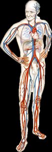
The cardiovascular system is responsible for transporting blood, nutrients, gases, and wastes throughout the body. It consists of the heart, blood vessels, and blood. The heart acts as a pump, propelling blood through a closed system of vessels.
Chapter 18: The Heart
Anatomical Location of the Heart
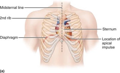
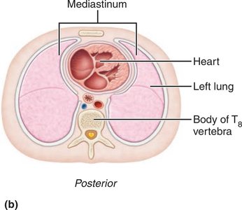
The heart is located in the mediastinum, the central compartment of the thoracic cavity, between the sternum and vertebral column, and between the lungs. It rests on the diaphragm and is bordered superiorly by the first rib.
Cardiopulmonary Resuscitation (CPR)
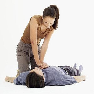
CPR is a life-saving technique that combines chest compressions with artificial ventilation to maintain circulatory flow and oxygenation during cardiac arrest. The heart's position between the sternum and vertebral column allows effective compressions to propel blood.
Layers of the Heart Wall
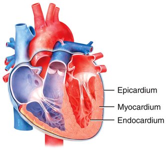
The heart wall consists of three layers:
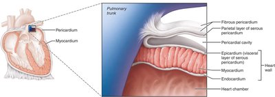
Epicardium: The outermost layer, also known as the visceral layer of the serous pericardium.
Myocardium: The thick, muscular middle layer responsible for contraction.
Endocardium: The innermost layer lining the heart chambers and valves.
Pericardium and Pericardial Disorders
The pericardium is a double-walled sac that surrounds and protects the heart. It consists of the fibrous pericardium (outer) and serous pericardium (inner). Inflammation of the pericardium is called pericarditis, which can lead to cardiac tamponade, a dangerous accumulation of fluid that restricts heart expansion.
Inflammation of the Heart
Myocarditis: Inflammation of the myocardium, often due to viral infection, rheumatic fever, or toxins.
Endocarditis: Inflammation of the endocardium, usually caused by bacterial infection, affecting heart valves.
Chambers of the Heart
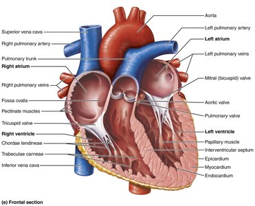
The heart has four chambers:
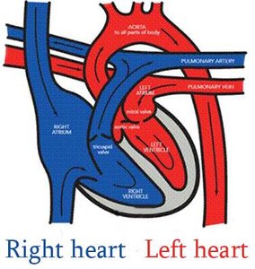
Right atrium and right ventricle: Receive deoxygenated blood from the body and pump it to the lungs (pulmonary circulation).
Left atrium and left ventricle: Receive oxygenated blood from the lungs and pump it to the body (systemic circulation).
Internal Anatomy of the Heart
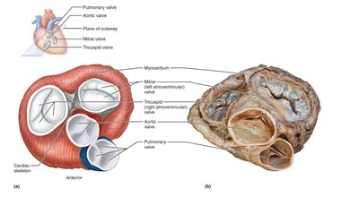
The heart contains valves that ensure unidirectional blood flow:
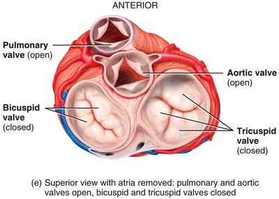
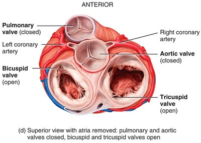
Atrioventricular (AV) valves: Tricuspid (right) and bicuspid/mitral (left) valves, between atria and ventricles.
Semilunar valves: Pulmonary and aortic valves, between ventricles and major arteries.
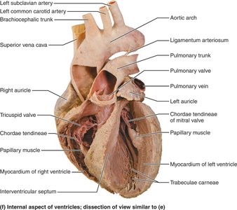
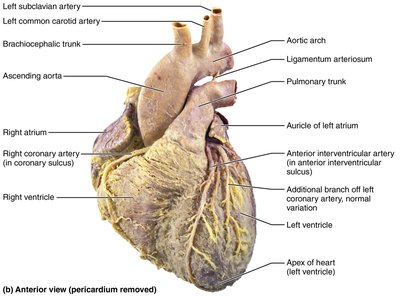
Comparison of Heart Chambers and Ventricles
The atria are thin-walled and act as receiving chambers, while the ventricles are thick-walled and serve as the main pumps. The left ventricle is thicker than the right because it must generate higher pressure to pump blood throughout the body.
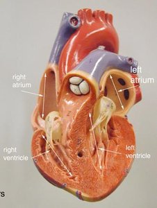
Heart Valves: Structure and Function
Valves open and close in response to pressure differences, preventing backflow:
AV valves prevent backflow into atria during ventricular contraction.
Semilunar valves prevent backflow into ventricles after ejection.
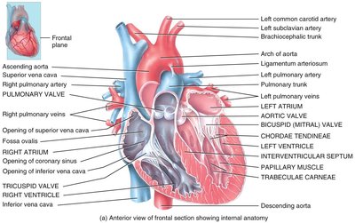
Cardiac Cycle
The cardiac cycle includes all events in one heartbeat:
Systole: Contraction phase (ventricular or atrial).
Diastole: Relaxation phase (ventricular or atrial).
Blood Flow Through the Heart
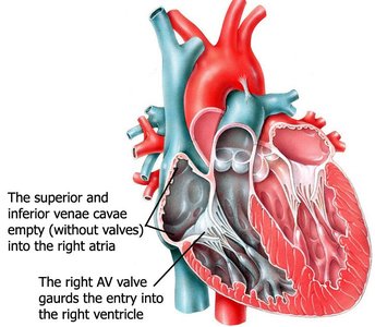
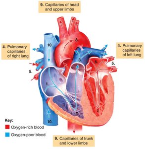
Blood flows through the heart in a specific sequence, ensuring oxygenation and systemic delivery:
Deoxygenated blood enters the right atrium via the superior and inferior vena cava.
Passes through the tricuspid valve to the right ventricle.
Pumped through the pulmonary valve into the pulmonary trunk and arteries to the lungs.
Oxygenated blood returns via pulmonary veins to the left atrium.
Passes through the mitral valve to the left ventricle.
Pumped through the aortic valve into the aorta and systemic circulation.
Coronary Circulation
The myocardium receives oxygen and nutrients from coronary arteries, which branch off the aorta. Venous blood from the myocardium returns to the right atrium via the coronary sinus.
Myocardial Ischemia and Infarction
Ischemia: Reduced blood flow to the myocardium, causing angina pectoris (chest pain).
Myocardial infarction (heart attack): Prolonged ischemia leading to tissue death and scar formation.
Histology of Cardiac Muscle
Cardiac muscle fibers are striated, branched, and interconnected by intercalated discs containing desmosomes and gap junctions. They have abundant mitochondria and rely on aerobic respiration.
Cardiac Conduction System
The heart's electrical system ensures coordinated contraction:
Sinoatrial (SA) node: Pacemaker, initiates impulse.
Atrioventricular (AV) node: Delays impulse, allowing atrial contraction.
AV bundle (Bundle of His): Conducts impulse to ventricles.
Right and left bundle branches: Carry impulse through interventricular septum.
Purkinje fibers: Distribute impulse to ventricular myocardium.
Electrocardiogram (ECG/EKG)
An ECG records the electrical activity of the heart. Key waves include:
P wave: Atrial depolarization
QRS complex: Ventricular depolarization (and atrial repolarization)
T wave: Ventricular repolarization
Heart Sounds
Heart sounds are produced by valve closures:
Lub (S1): Closure of AV valves
Dub (S2): Closure of semilunar valves
Cardiac Output (CO)
Cardiac output is the volume of blood ejected by the left ventricle per minute:
CO = Stroke Volume (SV) × Heart Rate (HR)
For example, if SV = 70 mL/beat and HR = 75 beats/min:
Regulation of Cardiac Output
Preload: Degree of stretch of the heart before contraction (Frank-Starling Law).
Contractility: Strength of contraction at a given preload.
Afterload: Pressure that must be overcome to eject blood.
Chapter 19: Blood Vessels and Hemodynamics
Types of Blood Vessels
Arteries: Carry blood away from the heart; thick-walled and elastic.
Arterioles: Small arteries that regulate blood flow into capillaries.
Capillaries: Site of exchange of gases, nutrients, and wastes.
Venules: Collect blood from capillaries.
Veins: Return blood to the heart; contain valves to prevent backflow.
Hemodynamics: Blood Flow, Pressure, and Resistance
Blood flow is determined by the pressure gradient and resistance:
F: Flow (cardiac output)
P: Pressure difference
R: Resistance
Blood pressure is the force exerted by blood on vessel walls, measured in mmHg. Mean arterial pressure (MAP) is calculated as:
Factors Affecting Resistance
Vessel radius: Resistance is inversely proportional to the fourth power of the radius ().
Blood viscosity: Increased viscosity increases resistance.
Vessel length: Longer vessels increase resistance.
Vasoconstriction increases resistance and decreases flow; vasodilation decreases resistance and increases flow.
Venous Return
Venous return is aided by pressure gradients, venous valves, skeletal muscle pump, and respiratory pump. Venous return must equal cardiac output for effective circulation.
Capillary Exchange
Diffusion: Passive movement of substances along concentration gradients.
Bulk flow: Movement of large volumes of fluid driven by pressure differences (Starling forces).
Starling's Law describes the balance between filtration and reabsorption at the capillary level.
Control of Blood Pressure and Blood Flow
Baroreceptor reflexes: Detect changes in blood pressure and adjust heart rate and vessel diameter.
Hormonal regulation: Includes the renin-angiotensin-aldosterone system, epinephrine/norepinephrine, antidiuretic hormone, and atrial natriuretic peptide.
Autoregulation: Local control of blood flow to match tissue metabolic needs via myogenic and metabolic responses.
Summary Table: Types of Blood Vessels
Vessel Type | Main Function | Key Features |
|---|---|---|
Arteries | Carry blood away from heart | Thick, elastic walls |
Arterioles | Regulate blood flow to capillaries | Thick smooth muscle layer |
Capillaries | Exchange of gases/nutrients | Thin walls, single cell layer |
Venules | Collect blood from capillaries | Thin walls |
Veins | Return blood to heart | Valves, thin walls |
Additional info: This guide integrates foundational concepts from Chapters 18 and 19, including heart anatomy, physiology, and vascular function, as required for ANP college-level study.
