 Back
BackThe Cardiovascular System: The Heart and Blood Vessels
Study Guide - Smart Notes
 Tailored notes based on your materials, expanded with key definitions, examples, and context.
The Structure and Function of the Heart
Anatomy of the Heart
The heart is a muscular organ responsible for pumping blood throughout the body via the circulatory system. It consists of four chambers: two atria and two ventricles, separated by valves that ensure unidirectional blood flow. The walls of the heart are composed of three layers: epicardium (outer), myocardium (middle, muscular), and endocardium (inner, endothelial).
Epicardium: The external surface, also known as the visceral pericardium.
Myocardium: Contains cardiac muscle cells, connective tissue, blood vessels, and nerves.
Endocardium: The internal endothelial surface.
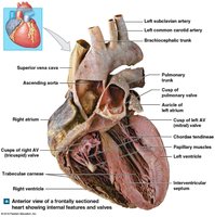
Heart Chambers and Valves
The heart is divided into right and left sides, each with an atrium and a ventricle. The right side receives deoxygenated blood and pumps it to the lungs (pulmonary circulation), while the left side receives oxygenated blood and pumps it to the body (systemic circulation).
Atria: Thin-walled chambers that receive blood.
Ventricles: Thick-walled chambers that pump blood out of the heart.
Valves:
Atrioventricular (AV) valves: Tricuspid (right) and bicuspid/mitral (left) valves.
Semilunar valves: Pulmonary (right) and aortic (left) valves.
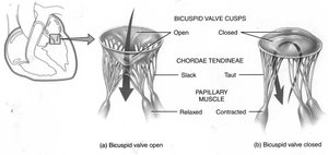
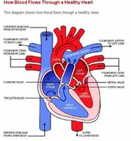
Blood Flow Through the Heart
Pathway of Blood
Blood flows through the heart in a specific sequence, passing through each chamber and valve.
Right atrium
Tricuspid valve
Right ventricle
Pulmonary semilunar valve
Pulmonary trunk and arteries
Lungs (gas exchange)
Pulmonary veins
Left atrium
Bicuspid (mitral) valve
Left ventricle
Aortic semilunar valve
Aorta
Body tissues
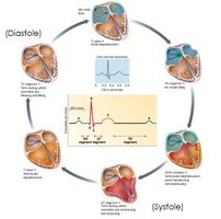
The Cardiac Cycle
Phases of the Cardiac Cycle
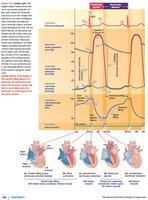
The cardiac cycle consists of alternating periods of contraction (systole) and relaxation (diastole) of the heart chambers.
Ventricular filling: Blood flows passively into the ventricles during diastole.
Isovolumetric contraction: Ventricles contract with all valves closed, building pressure.
Ventricular ejection: Semilunar valves open, blood is pumped out.
Isovolumetric relaxation: Ventricles relax, all valves closed, pressure drops.
Pressure and Volume Changes
Pressure changes in the chambers and vessels drive the opening and closing of valves, ensuring efficient blood flow.
AV valves open when atrial pressure exceeds ventricular pressure.
Semilunar valves open when ventricular pressure exceeds vessel pressure.
Heart sounds ("lub-dub") are produced by valve closure.
Electrical Activity of the Heart
Intrinsic Conduction System
The heart's electrical activity is generated and propagated by specialized cells.
SA node: Pacemaker cells initiate action potentials.
AV node: Delays impulse, allowing atrial contraction.
Bundle of His and Purkinje fibers: Rapidly conduct impulses to ventricles.
Electrocardiogram (ECG/EKG)
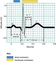
An ECG records the electrical events of the heart.
P wave: Atrial depolarization
QRS complex: Ventricular depolarization
T wave: Ventricular repolarization
Cardiac Muscle Action Potentials
Phases of Action Potential in Cardiac Contractile Cells
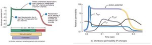
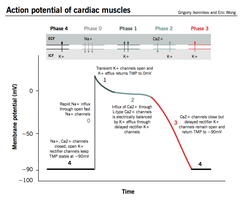
Cardiac muscle cells exhibit a unique action potential with a plateau phase, preventing tetanus and allowing proper contraction.
Phase 0: Rapid depolarization due to Na+ influx
Phase 1: Partial repolarization due to transient K+ efflux
Phase 2: Plateau phase due to Ca2+ influx
Phase 3: Repolarization due to K+ efflux
Phase 4: Resting membrane potential
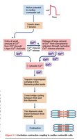
Excitation-Contraction Coupling
The process by which electrical signals trigger muscle contraction involves Ca2+ influx and release from the sarcoplasmic reticulum, leading to crossbridge cycling.
Ca2+ binds to troponin, moving tropomyosin and allowing actin-myosin interaction.
Extent of contraction depends on cytosolic Ca2+ concentration.
Action Potential of Autorhythmic Cells
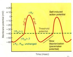
Pacemaker Potential
Autorhythmic cells (e.g., SA node) generate spontaneous action potentials due to slow Na+ and Ca2+ influx and reduced K+ efflux.
Threshold reached at -40 mV, Ca2+ channels open for rapid depolarization.
Repolarization occurs as Ca2+ channels close and K+ channels open.
Regulation of Heart Rate and Cardiac Output
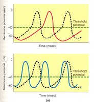
Autonomic Nervous System Control
The heart rate is regulated by the autonomic nervous system (ANS):
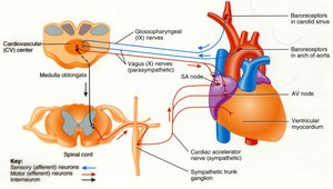
Parasympathetic (vagus nerve): Releases ACh, opens K+ channels, slows depolarization, decreases heart rate.
Sympathetic: Releases norepinephrine, closes K+ channels, increases Ca2+ influx, speeds depolarization, increases heart rate.
Cardiac Output
Cardiac output (CO) is the volume of blood pumped by each ventricle per minute.
Formula:
Heart Rate (HR): Beats per minute
Stroke Volume (SV): Volume of blood pumped per beat
Blood Pressure Regulation
Factors Affecting Blood Pressure
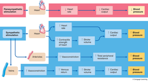
Blood pressure is determined by cardiac output and peripheral resistance.
Increase in output or resistance raises blood pressure.
Decrease in output or resistance lowers blood pressure.
Blood volume, vessel diameter, and viscosity also play roles.
Local and Extrinsic Control of Blood Flow
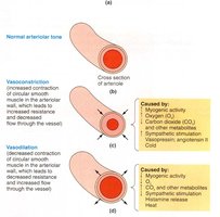
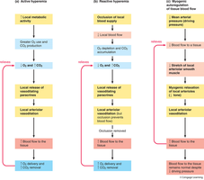
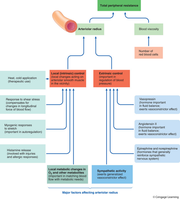
Local factors: Metabolic changes, histamine, myogenic activity, shear stress, temperature.
Extrinsic factors: Sympathetic stimulation, hormones (norepinephrine, epinephrine, vasopressin, angiotensin II).
Baroreceptor Reflex and Blood Pressure Homeostasis
Baroreceptor Reflex
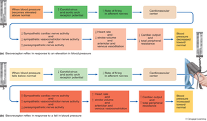
Baroreceptors in the aorta and carotid sinus monitor blood pressure and send signals to the cardiovascular center in the medulla.
Increased pressure decreases sympathetic activity, causing vasodilation and reduced heart rate.
Decreased pressure increases sympathetic activity, causing vasoconstriction and increased heart rate.
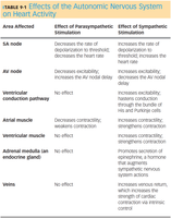
Effects of the Autonomic Nervous System on Heart Activity
Comparison Table
Area Affected | Parasympathetic Effect | Sympathetic Effect |
|---|---|---|
SA node | Decreases rate of depolarization, decreases heart rate | Increases rate of depolarization, increases heart rate |
AV node | Decreases excitability | Increases excitability |
Ventricular conduction pathway | No effect | Increases conduction velocity |
Atrial muscle | Weakens contraction | Strengthens contraction |
Adrenal medulla | No effect | Promotes secretion of epinephrine |
Veins | No effect | Increases vasoconstriction |
Summary of ANS Effects on Blood Pressure
Elastic Properties of Arteries
Role of Elastic Arteries
The aorta and elastic arteries stretch during ventricular contraction and recoil during relaxation, helping maintain continuous blood flow. 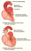
Pressure Changes in the Circulatory System
Blood Pressure Gradient
Blood pressure is highest in the arteries and decreases as blood moves through arterioles, capillaries, venules, and veins. 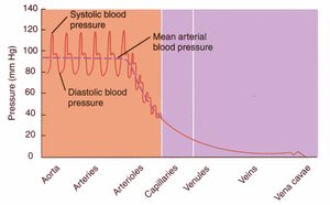
Additional info:
Cardiac muscle cells are striated, branched, and possess intercalated discs for rapid electrical transmission.
Cardiac output and blood pressure are tightly regulated by neural and hormonal mechanisms.
Vasovagal response is a rapid decrease in blood pressure due to increased parasympathetic stimulation, leading to fainting.
