 Back
BackThe Cardiovascular System: The Heart – Structure, Function, and Clinical Correlates
Study Guide - Smart Notes
 Tailored notes based on your materials, expanded with key definitions, examples, and context.
The Cardiovascular System: The Heart
Location and Basic Structure of the Heart
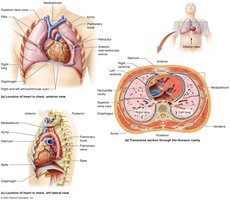
The heart is a muscular organ located in the thoracic cavity, between the lungs in the mediastinum. It is slightly tilted so that the apex points to the left hip and the base is directed toward the right shoulder. The heart is divided into four chambers: two atria (upper chambers) and two ventricles (lower chambers). The right side of the heart pumps blood to the lungs (pulmonary circuit), while the left side pumps blood to the rest of the body (systemic circuit).
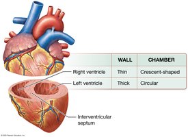
Right Ventricle: Thin-walled, crescent-shaped chamber that pumps blood to the lungs.
Left Ventricle: Thick-walled, circular chamber that pumps blood to the systemic circuit.
Functions of the Heart
The heart acts as a double pump, maintaining two separate circuits:
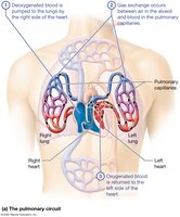
Pulmonary Circuit: Carries deoxygenated blood from the right ventricle to the lungs for gas exchange and returns oxygenated blood to the left atrium.
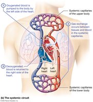
Systemic Circuit: Delivers oxygenated blood from the left ventricle to the body tissues and returns deoxygenated blood to the right atrium.
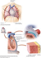
The Pericardium and Heart Wall
The Pericardium
The pericardium is a double-walled sac that surrounds and protects the heart. It consists of two main layers:
Fibrous Pericardium: Tough, outer layer that anchors the heart and prevents overfilling.
Serous Pericardium: Thin, inner layer with two sublayers (parietal and visceral) separated by serous fluid, reducing friction during heartbeats.
Layers of the Heart Wall
The heart wall is composed of three layers:
Epicardium (Visceral Pericardium): Outermost layer, provides protection and contains blood vessels.
Myocardium: Middle, muscular layer responsible for contraction and pumping action.
Endocardium: Innermost layer, lines the chambers and valves, minimizing friction.
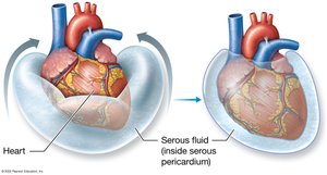
Cardiac Tamponade
Cardiac tamponade is a clinical condition where excess fluid accumulates in the pericardial cavity, compressing the heart and reducing its ability to fill and pump blood. Causes include trauma, cancer, kidney failure, or surgery. Treatment involves removing the fluid via pericardiocentesis.

External and Internal Anatomy of the Heart
External Anatomy
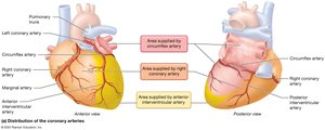
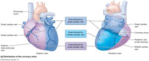
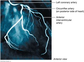
The heart's external features include the auricles, major vessels (aorta, pulmonary arteries and veins, vena cavae), and coronary vessels. The coronary arteries supply the heart muscle with oxygenated blood.
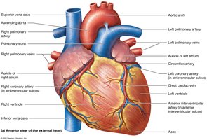
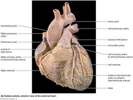
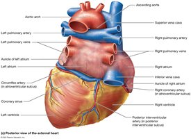
Internal Anatomy
Internally, the heart is divided by the interventricular septum and contains valves that ensure unidirectional blood flow. The right atrium receives blood from the body, and the left atrium receives blood from the lungs. The ventricles pump blood to the pulmonary and systemic circuits.
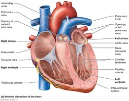
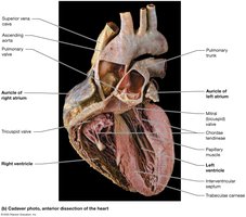
Heart Valves and Valvular Diseases
Heart Valves
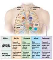
The heart contains four main valves:
Atrioventricular (AV) Valves: Tricuspid (right) and mitral (left) valves, prevent backflow into the atria during ventricular contraction.
Semilunar Valves: Pulmonary and aortic valves, prevent backflow into the ventricles after contraction.
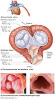
Valvular Heart Diseases
Valvular defects can be congenital or acquired (infection, cancer, immune disorders):
Insufficient Valve (Regurgitation): Valve fails to close fully, causing blood to leak backward.
Stenotic Valve: Valve cusps become stiff and inflexible, restricting blood flow.

Heart Sounds
Normal heart sounds (S1 "lub" and S2 "dub") are produced by valve closures. Abnormal sounds (murmurs) indicate defective valves.
Coronary Circulation and Disease
Coronary Circulation
The coronary arteries supply oxygenated blood to the myocardium, while coronary veins drain deoxygenated blood. Blockage of these vessels can lead to myocardial infarction (heart attack).
Coronary Artery Disease (CAD)
CAD is caused by atherosclerosis (plaque buildup) in the coronary arteries, reducing blood flow to the heart muscle. Risk factors include high cholesterol, hypertension, smoking, and diabetes. Symptoms may include chest pain (angina) or myocardial infarction.
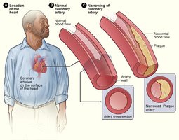
Treatment of CAD
Treatment options include lifestyle changes, medications, angioplasty with stent placement, and coronary artery bypass grafting (CABG).


Cardiac Muscle Tissue and Electrophysiology
Cardiac Muscle Tissue
Cardiac muscle exhibits autorhythmicity, meaning it can generate its own action potentials without nervous input. There are two main cell types:
Pacemaker Cells: Generate rhythmic action potentials to set the heart rate.
Contractile Cells: Respond to pacemaker signals and contract to pump blood.
Cardiac Electrophysiology
The heart's electrical activity is coordinated by the cardiac conduction system, including the SA node (primary pacemaker), AV node, AV bundle, bundle branches, and Purkinje fibers. The SA node sets the normal sinus rhythm. Action potentials in contractile cells feature a plateau phase, which prolongs contraction and prevents tetany.
Electrocardiogram (ECG)
An ECG records the electrical activity of the heart, providing information about heart rhythm and conduction. Common abnormalities include bradycardia, tachycardia, heart block, and fibrillation.
The Cardiac Cycle and Cardiac Output
The Cardiac Cycle
The cardiac cycle is the sequence of events in one heartbeat, including atrial and ventricular systole (contraction) and diastole (relaxation). Pressure changes drive blood flow and valve operation.
Stroke Volume and Cardiac Output
Stroke Volume (SV): The amount of blood ejected by a ventricle in one contraction. Calculated as:
where EDV = end-diastolic volume, ESV = end-systolic volume.
Cardiac Output (CO): The volume of blood pumped by each ventricle per minute. Calculated as:
where HR = heart rate (beats per minute).
Regulation of Cardiac Output
Cardiac output is regulated by preload (venous return), contractility (strength of contraction), and afterload (resistance in arteries). The autonomic nervous system and hormones also influence heart rate and stroke volume.
Heart Failure
Heart failure occurs when the heart cannot pump blood effectively. Causes include myocardial infarction, valvular disease, cardiomyopathy, and ventricular hypertrophy. Left-sided failure leads to pulmonary edema, while right-sided failure causes systemic edema. Treatment focuses on improving cardiac output and reducing fluid overload.
Additional info: For a deeper understanding, students should review the mechanisms of action potentials in cardiac muscle, the phases of the cardiac cycle, and the clinical significance of ECG waveforms.
