 Back
BackThe Cardiovascular System: The Heart – Structure, Function, and Regulation
Study Guide - Smart Notes
 Tailored notes based on your materials, expanded with key definitions, examples, and context.
The Cardiovascular System: The Heart
Overview of the Heart and Circulatory Pathways
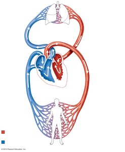
The heart is a muscular organ responsible for pumping blood throughout the body via two main circuits: the pulmonary and systemic circuits. It ensures the delivery of oxygen and nutrients to tissues and the removal of carbon dioxide and metabolic wastes.
Pulmonary Circuit: Carries deoxygenated blood from the right side of the heart to the lungs for gas exchange, then returns oxygenated blood to the left side of the heart.
Systemic Circuit: Distributes oxygen-rich blood from the left side of the heart to body tissues and returns deoxygenated blood to the right side.
Location and Gross Anatomy of the Heart
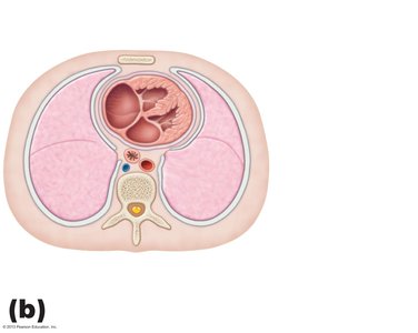
The heart is located in the mediastinum, between the lungs, posterior to the sternum, and superior to the diaphragm. Its apex points to the left hip, and its base is directed toward the right shoulder.
Mediastinum: Central compartment of the thoracic cavity containing the heart, great vessels, and other structures.
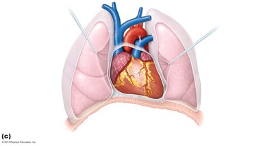
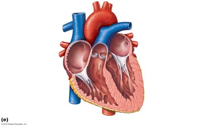
Chambers: The heart has four chambers: two atria (superior) and two ventricles (inferior).
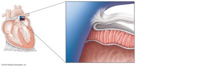
Heart Wall and Pericardium
The heart wall consists of three layers: epicardium, myocardium, and endocardium. The heart is enclosed in a double-walled sac called the pericardium, which protects and anchors the heart.
Fibrous Pericardium: Tough outer layer that prevents overfilling.
Serous Pericardium: Double-layered membrane (parietal and visceral layers) with a lubricating pericardial cavity between them.
Myocardium: Cardiac muscle layer responsible for contraction.
Endocardium: Endothelial lining continuous with blood vessels.
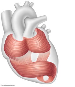
Microscopic Anatomy of Cardiac Muscle
Cardiac muscle cells are striated, branched, and interconnected by intercalated discs containing gap junctions and desmosomes. This structure allows for coordinated contraction and efficient pumping.
Intercalated Discs: Specialized connections that facilitate electrical and mechanical coupling between cells.
Myocardial Bundles: Arranged in spiral and circular patterns to optimize blood ejection.
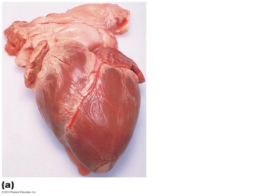
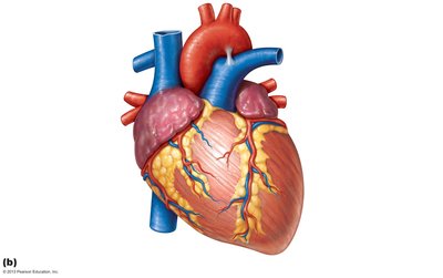
Gross Anatomy: External and Internal Features
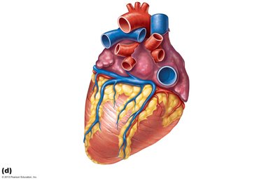
The heart's external anatomy includes major vessels, coronary arteries, and veins. Internally, the heart contains valves that ensure unidirectional blood flow and prevent backflow.
Major Vessels: Aorta, pulmonary trunk, venae cavae, pulmonary veins.
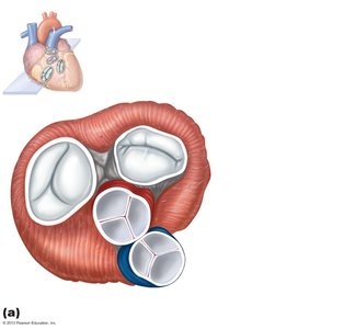
Valves: Atrioventricular (tricuspid and mitral) and semilunar (aortic and pulmonary) valves.
Heart Valves and Blood Flow
Heart valves maintain unidirectional blood flow through the heart. The atrioventricular (AV) valves prevent backflow into the atria, while the semilunar (SL) valves prevent backflow into the ventricles.
AV Valves: Tricuspid (right), Mitral/Bicuspid (left)
SL Valves: Pulmonary (right), Aortic (left)
Chordae Tendineae and Papillary Muscles: Prevent valve prolapse during ventricular contraction.
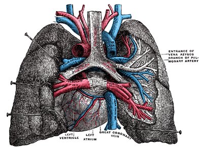
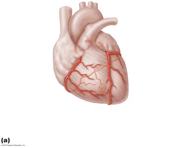
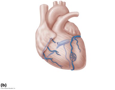
Coronary Circulation
The heart muscle (myocardium) receives its blood supply from the coronary arteries. Venous blood is collected by cardiac veins and returned to the right atrium via the coronary sinus.
Coronary Arteries: Right and left coronary arteries, with major branches supplying the heart wall.
Cardiac Veins: Great, middle, and small cardiac veins drain into the coronary sinus.
Anastomoses: Provide collateral circulation in case of arterial blockage.
Cardiac Muscle Physiology and Contraction
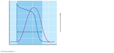
Cardiac muscle cells are self-excitable (automaticity), contract as a unit, and have a long refractory period to prevent tetanus. The action potential in cardiac muscle involves rapid depolarization, a plateau phase, and repolarization.
Automaticity: Pacemaker cells spontaneously depolarize to initiate each heartbeat.
Action Potential Phases: Depolarization (Na+ influx), Plateau (Ca2+ influx), Repolarization (K+ efflux).
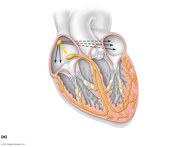
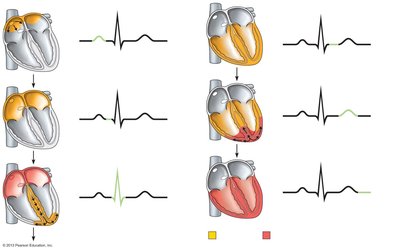
Intrinsic Conduction System
The intrinsic conduction system coordinates the heart's electrical activity, ensuring efficient contraction. Key components include the SA node, AV node, AV bundle, bundle branches, and Purkinje fibers.
SA Node: Primary pacemaker, initiates heartbeat.
AV Node: Delays impulse, allowing atrial contraction before ventricular contraction.
AV Bundle and Branches: Conduct impulses to ventricles.
Purkinje Fibers: Distribute impulse through ventricular myocardium.
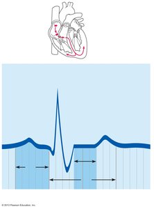
Electrocardiogram (ECG) and Cardiac Cycle
An ECG records the electrical activity of the heart. The main waves are the P wave (atrial depolarization), QRS complex (ventricular depolarization), and T wave (ventricular repolarization). The cardiac cycle includes all events associated with one heartbeat.
P Wave: Atrial depolarization
QRS Complex: Ventricular depolarization
T Wave: Ventricular repolarization
Cardiac Cycle Phases: Atrial systole, ventricular systole, and diastole
Cardiac Output and Its Regulation
Cardiac output (CO) is the volume of blood pumped by each ventricle per minute. It is determined by heart rate (HR) and stroke volume (SV):
Formula:
Stroke Volume (SV): Difference between end-diastolic volume (EDV) and end-systolic volume (ESV):
Regulation: Preload (venous return), contractility, and afterload (arterial pressure) affect SV. Autonomic nervous system and hormones regulate HR.
Congestive Heart Failure and Clinical Correlations
Congestive heart failure (CHF) occurs when the heart cannot pump sufficient blood to meet tissue needs. Causes include coronary artery disease, hypertension, myocardial infarction, valve defects, and cardiomyopathies. Left-sided failure leads to pulmonary congestion; right-sided failure causes systemic edema.
Common Causes: Atherosclerosis, chronic hypertension, myocardial infarction, valve defects, emphysema.
Clinical Signs: Edema, shortness of breath, fatigue.
Developmental Aspects and Congenital Heart Defects
The heart develops from a simple tube that folds and partitions to form four chambers. Congenital heart defects are structural abnormalities present at birth, such as ventricular septal defect, coarctation of the aorta, and tetralogy of Fallot.
Ventricular Septal Defect: Opening in the interventricular septum allows blood mixing.
Coarctation of the Aorta: Narrowing of the aorta increases left ventricular workload.
Tetralogy of Fallot: Combination of four defects affecting blood flow and oxygenation.
Defect | Description | Incidence |
|---|---|---|
Ventricular Septal Defect | Superior part of interventricular septum fails to form; blood mixes between ventricles | 1 in 500 births |
Coarctation of the Aorta | Part of aorta is narrowed; increases left ventricular workload | 1 in 1500 births |
Tetralogy of Fallot | Four defects: pulmonary stenosis, right ventricular hypertrophy, ventricular septal defect, overriding aorta | 1 in 2000 births |
