 Back
The Cardiovascular System: The Heart – Structure, Function, and Regulation
Study Guide - Smart Notes
 Tailored notes based on your materials, expanded with key definitions, examples, and context.
The Cardiovascular System: The Heart
Overview of Heart Function and Circuits
The heart is a muscular organ that serves as the central pump of the cardiovascular system, ensuring the circulation of blood throughout the body. It operates as two side-by-side pumps, each serving a distinct circuit:
Pulmonary Circuit: The right side of the heart receives oxygen-poor blood from the body and pumps it to the lungs for gas exchange (removal of CO2 and uptake of O2).
Systemic Circuit: The left side receives oxygen-rich blood from the lungs and pumps it to the rest of the body tissues.
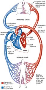
Additional info: The heart ensures unidirectional blood flow and separates oxygenated and deoxygenated blood, which is essential for efficient metabolic support of tissues.
Heart Anatomy
Size, Location, and Orientation
The heart is approximately the size of a fist and weighs less than 1 pound. It is located in the mediastinum, between the second rib and fifth intercostal space, resting on the superior surface of the diaphragm. Two-thirds of the heart lies to the left of the midsternal line, anterior to the vertebral column and posterior to the sternum.
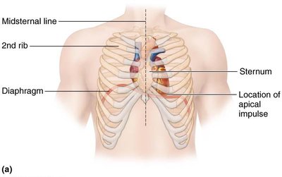
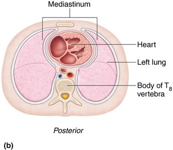
Base: Leans toward the right shoulder.
Apex: Points toward the left hip; the apical impulse can be palpated between the fifth and sixth ribs, just below the left nipple.
Coverings of the Heart
The heart is enclosed in a double-walled sac called the pericardium:
Fibrous pericardium: Superficial layer that protects, anchors, and prevents overfilling.
Serous pericardium: Deep, two-layered membrane (parietal and visceral layers) separated by the pericardial cavity, which contains fluid to reduce friction.
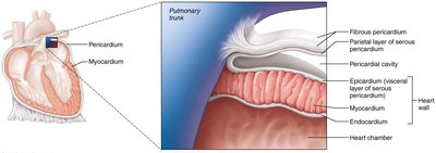
Clinical Note: Pericarditis is inflammation of the pericardium, which can lead to pericardial friction rub and, in severe cases, cardiac tamponade (compression of the heart by fluid).
Layers of the Heart Wall
The heart wall consists of three layers:
Epicardium: Visceral layer of the serous pericardium.
Myocardium: Cardiac muscle layer responsible for contraction; contains the cardiac skeleton (connective tissue network).
Endocardium: Endothelial layer lining the heart chambers and valves.
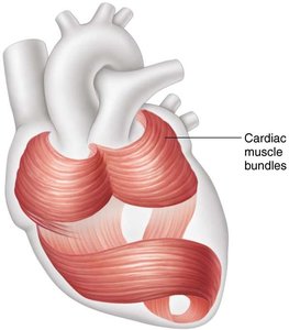
Chambers and Associated Great Vessels
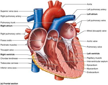
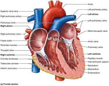
The heart has four chambers:
Atria (right and left): Superior, thin-walled receiving chambers.
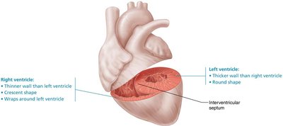
Ventricles (right and left): Inferior, thick-walled pumping chambers.
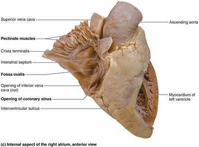
Interatrial septum: Separates the atria; contains the fossa ovalis (remnant of fetal foramen ovale).
Interventricular septum: Separates the ventricles.
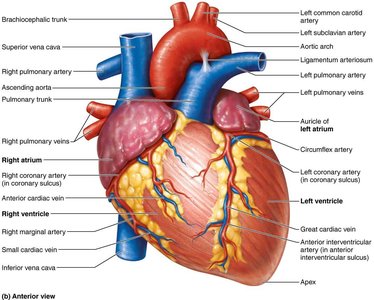
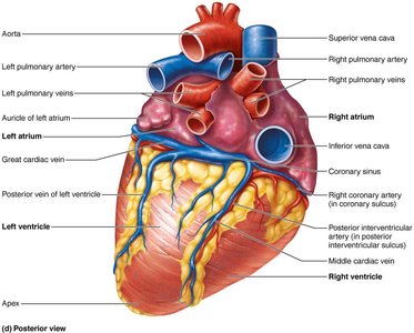
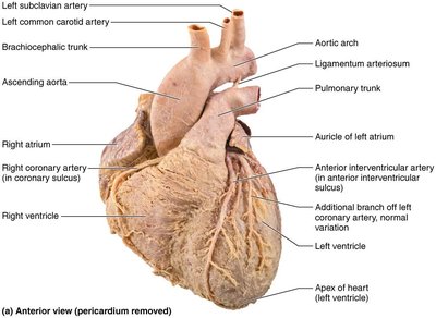
Surface features include the coronary sulcus (atrioventricular groove), anterior and posterior interventricular sulci, which mark the boundaries between chambers.
Atria: The Receiving Chambers
Right atrium: Receives deoxygenated blood from the superior vena cava, inferior vena cava, and coronary sinus.
Left atrium: Receives oxygenated blood from four pulmonary veins.
Auricles: Small appendages that increase atrial volume.
Ventricles: The Discharging Chambers
Right ventricle: Pumps blood into the pulmonary trunk (to the lungs).
Left ventricle: Pumps blood into the aorta (to the systemic circuit); has thicker walls due to higher pressure workload.
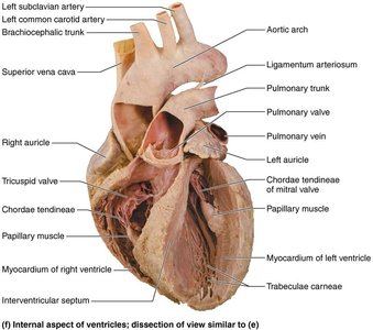
Trabeculae carneae: Irregular muscle ridges on ventricular walls.
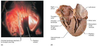
Papillary muscles: Anchor chordae tendineae attached to heart valves.
Heart Valves
Types and Functions of Heart Valves
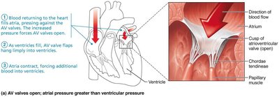
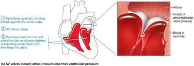
Heart valves ensure unidirectional blood flow and open/close in response to pressure changes:
Atrioventricular (AV) valves: Between atria and ventricles (tricuspid on right, mitral/bicuspid on left).
Semilunar (SL) valves: Between ventricles and major arteries (pulmonary and aortic valves).
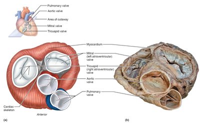
Atrioventricular (AV) Valves
Tricuspid valve: Right AV valve, three cusps.
Mitral (bicuspid) valve: Left AV valve, two cusps.
Chordae tendineae: Anchor valve cusps to papillary muscles, preventing eversion.
Semilunar (SL) Valves
Pulmonary SL valve: Between right ventricle and pulmonary trunk.
Aortic SL valve: Between left ventricle and aorta.
Each has three half-moon-shaped cusps.
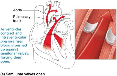
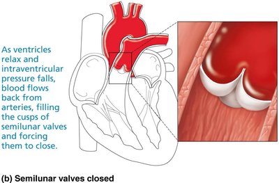
Clinical Note: Valve disorders include incompetent valves (backflow) and valvular stenosis (narrowing), which may require surgical replacement.
Pathway of Blood Through the Heart
Right Side (Pulmonary Circuit)
Superior/inferior vena cava and coronary sinus →
Right atrium →
Tricuspid valve →
Right ventricle →
Pulmonary semilunar valve →
Pulmonary trunk →
Pulmonary arteries →
Lungs
Left Side (Systemic Circuit)
Four pulmonary veins →
Left atrium →
Mitral valve →
Left ventricle →
Aortic semilunar valve →
Aorta →
Systemic circulation
Equal volumes of blood are pumped by each side. The pulmonary circuit is low-pressure and short, while the systemic circuit is high-pressure and long. The left ventricle has a thicker wall to generate greater pressure.
Coronary Circulation
Arterial Supply
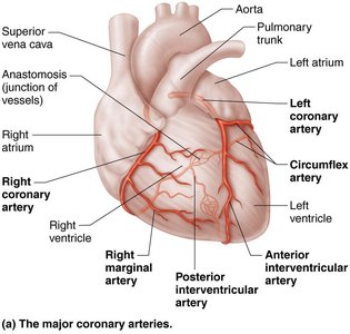
The heart muscle receives its own blood supply via the coronary arteries, which branch from the base of the aorta and encircle the heart in the coronary sulcus. Major branches include:
Left coronary artery: Anterior interventricular artery (LAD) and circumflex artery.
Right coronary artery: Right marginal artery and posterior interventricular artery.
Venous Drainage
Cardiac veins collect deoxygenated blood from the myocardium and drain into the coronary sinus, which empties into the right atrium. Major veins include the great cardiac vein, middle cardiac vein, and small cardiac vein.
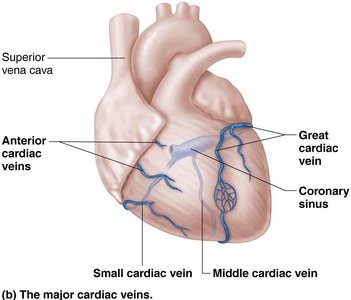
Clinical Note: Blockage of coronary arteries can cause angina pectoris or myocardial infarction (heart attack).
Microscopic Anatomy of Cardiac Muscle
Structure and Function
Cardiac muscle cells are striated, short, branched, and interconnected by intercalated discs.
Intercalated discs contain desmosomes (mechanical connection) and gap junctions (electrical connection), allowing the heart to function as a functional syncytium.
Cardiac muscle has abundant mitochondria and relies almost exclusively on aerobic respiration.
Additional info: The arrangement of sarcomeres and the presence of T tubules and a simpler sarcoplasmic reticulum distinguish cardiac from skeletal muscle.
Physiology of Cardiac Muscle Contraction
Comparison with Skeletal Muscle
Both types require depolarization and Ca2+ influx for contraction.
Cardiac muscle can be self-excitable (pacemaker cells), contracts as a unit, and has a longer refractory period, preventing tetanus.
Cardiac muscle relies on extracellular Ca2+ for contraction, unlike skeletal muscle.
Feature | Skeletal Muscle | Cardiac Muscle |
|---|---|---|
Contraction Initiation | Requires nervous stimulation | Can be self-excitable |
Contraction Duration | Short (1–2 ms AP) | Long (200 ms AP) |
Refractory Period | Short | Long (prevents tetanus) |
Energy Source | Aerobic/anaerobic | Primarily aerobic |
Additional info: The heart's reliance on aerobic metabolism makes it sensitive to oxygen deprivation.
Intrinsic Conduction System
Setting the Basic Rhythm
The heart's intrinsic conduction system consists of autorhythmic (pacemaker) cells that initiate and distribute electrical impulses, ensuring coordinated contraction:
Sinoatrial (SA) node (pacemaker) →
Atrioventricular (AV) node →
AV bundle (bundle of His) →
Right and left bundle branches →
Purkinje fibers (subendocardial conducting network)
Clinical Note: Defects in the conduction system can cause arrhythmias, fibrillation, or heart block, which may require interventions such as defibrillation or pacemakers.
Extrinsic Innervation
The autonomic nervous system (ANS) modulates heart rate and force:
Sympathetic stimulation: Increases heart rate and contractility.
Parasympathetic stimulation (vagus nerve): Decreases heart rate.
Action Potentials of Contractile Cardiac Muscle Cells
Cardiac muscle action potentials have a characteristic plateau phase due to prolonged Ca2+ influx, which ensures sustained contraction and efficient blood ejection.
Depolarization: Fast Na+ influx.
Plateau: Slow Ca2+ influx.
Repolarization: K+ efflux.
Equation:
Additional info: Longer refractory period prevents tetanic contractions, allowing the heart to relax and refill.
Electrocardiography (ECG/EKG)
An ECG records the electrical activity of the heart. Key features include:
P wave: Atrial depolarization
QRS complex: Ventricular depolarization (and atrial repolarization)
T wave: Ventricular repolarization
Intervals and segments (e.g., P-R, S-T, Q-T) provide diagnostic information about conduction and repolarization.
Mechanical Events of the Heart (Cardiac Cycle)
The cardiac cycle consists of systole (contraction) and diastole (relaxation) phases, coordinating blood flow through the heart:
Ventricular filling: AV valves open, blood flows into ventricles.
Isovolumetric contraction: All valves closed, ventricles contract, pressure rises.
Ventricular ejection: SL valves open, blood ejected into arteries.
Isovolumetric relaxation: All valves closed, ventricles relax.
Heart sounds: "Lub-dup" due to closing of AV and SL valves, respectively.
Clinical Note: Heart murmurs indicate abnormal valve function (incompetence or stenosis).
Regulation of Cardiac Output
Cardiac output (CO): Volume of blood pumped by each ventricle per minute.
Equation:
Stroke volume (SV): Volume of blood pumped per beat.
Heart rate (HR): Beats per minute.
Factors affecting SV:
Preload: Degree of stretch before contraction (Frank-Starling law).
Contractility: Strength of contraction at a given preload.
Afterload: Pressure ventricles must overcome to eject blood.
Factors affecting HR:
Autonomic nervous system (sympathetic/parasympathetic)
Hormones (epinephrine, thyroxine)
Ions (Ca2+, K+)
Other factors: age, gender, exercise, temperature
Clinical Note: Tachycardia (>100 bpm) and bradycardia (<60 bpm) can indicate underlying pathology or physiological adaptation.
Homeostatic Imbalances and Clinical Correlations
Congestive heart failure (CHF): Inadequate cardiac output due to weakened myocardium, often caused by coronary atherosclerosis, hypertension, myocardial infarcts, or dilated cardiomyopathy.
Pulmonary congestion: Left-sided failure; blood backs up in lungs.
Peripheral congestion: Right-sided failure; blood pools in body organs.
Developmental Aspects of the Heart
The heart develops from mesoderm, beginning as a simple tube that forms four chambers by day 35 of embryonic development. Fetal adaptations (foramen ovale, ductus arteriosus) bypass pulmonary circulation and close at birth.
Clinical Note: Congenital heart defects are the most common birth defects, often involving mixing of blood or narrowed vessels/valves (e.g., septal defects, patent ductus arteriosus, coarctation of the aorta, tetralogy of Fallot).
Age-related changes include valve sclerosis, decreased cardiac reserve, fibrosis of cardiac muscle, and atherosclerosis.
