 Back
The Cardiovascular System: The Heart – Structure, Function, and Physiology
Study Guide - Smart Notes
 Tailored notes based on your materials, expanded with key definitions, examples, and context.
The Cardiovascular System: The Heart
Overview of the Heart and Circulatory Circuits
The heart is a muscular organ responsible for pumping blood throughout the body via two main circuits: the pulmonary circuit and the systemic circuit. These circuits ensure the delivery of oxygen and nutrients to tissues and the removal of carbon dioxide and metabolic wastes.
Pulmonary Circuit: The right side of the heart receives oxygen-poor blood from the body and pumps it to the lungs for oxygenation.
Systemic Circuit: The left side of the heart receives oxygen-rich blood from the lungs and pumps it to the rest of the body.
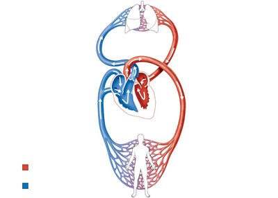
Key Point: The heart acts as two side-by-side pumps, each serving a different circuit.
Gross Anatomy of the Heart
Size, Location, and Orientation
The heart is approximately the size of a fist and is located in the mediastinum, between the second rib and the fifth intercostal space. About two-thirds of the heart lies to the left of the midsternal line, and it rests on the superior surface of the diaphragm.
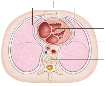
Base: The broad, superior aspect of the heart, directed toward the right shoulder.
Apex: The pointed, inferior aspect, directed toward the left hip.
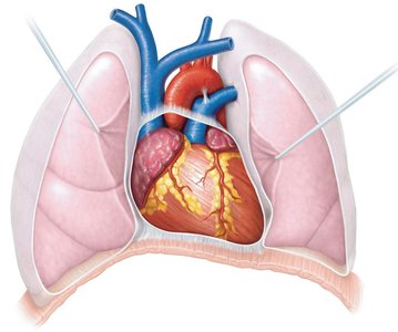
Coverings of the Heart: The Pericardium
The heart is enclosed in a double-walled sac called the pericardium, which consists of:
Fibrous pericardium: Tough, dense connective tissue that protects, anchors, and prevents overfilling of the heart.
Serous pericardium: Thin, double-layered membrane with a parietal layer lining the internal surface of the fibrous pericardium and a visceral layer (epicardium) covering the heart surface.
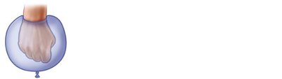
The pericardial cavity between these layers contains serous fluid to reduce friction during heart activity.
Layers of the Heart Wall
The heart wall is composed of three layers:
Epicardium: The outermost layer, also known as the visceral layer of the serous pericardium.
Myocardium: The thick, middle layer composed of cardiac muscle responsible for contraction.
Endocardium: The innermost layer lining the heart chambers and continuous with blood vessel endothelium.
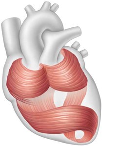
Cardiac skeleton: A network of connective tissue fibers within the myocardium that supports valves and vessels and limits the spread of electrical impulses.
Chambers of the Heart and Associated Vessels
Heart Chambers
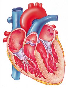
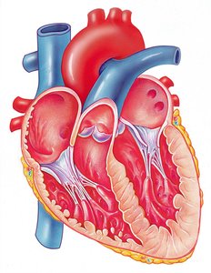
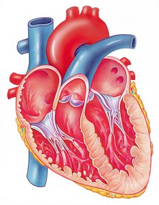
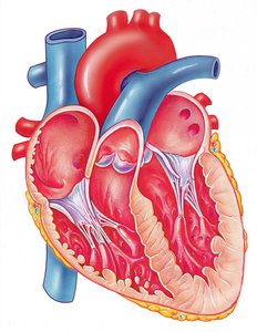
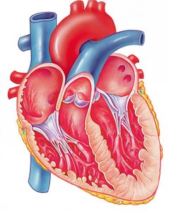
The heart contains four chambers:
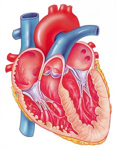
Two atria (right and left): Thin-walled receiving chambers separated by the interatrial septum.
Two ventricles (right and left): Thick-walled pumping chambers separated by the interventricular septum.
Major Vessels Entering and Leaving the Heart
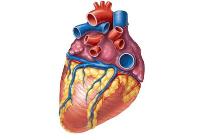
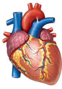
Right atrium: Receives deoxygenated blood from the superior vena cava, inferior vena cava, and coronary sinus.
Left atrium: Receives oxygenated blood from four pulmonary veins.
Right ventricle: Pumps blood into the pulmonary trunk (to the lungs).
Left ventricle: Pumps blood into the aorta (to the systemic circulation).
Heart Valves
Types and Functions of Heart Valves
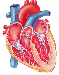
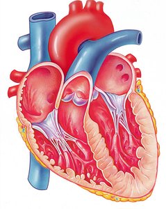
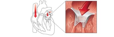
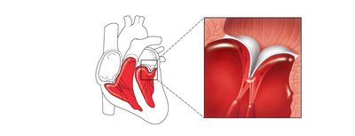
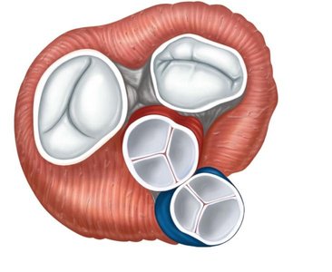
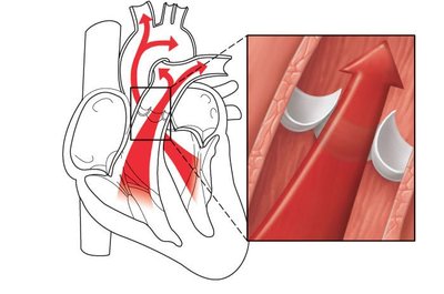
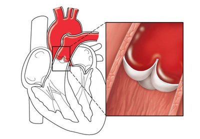
Heart valves ensure unidirectional blood flow through the heart by opening and closing in response to pressure changes. There are two main types:
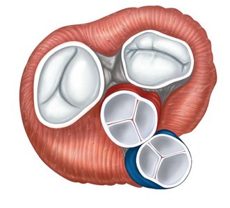
Atrioventricular (AV) valves: Located between atria and ventricles (tricuspid on right, mitral/bicuspid on left).
Semilunar (SL) valves: Located between ventricles and major arteries (pulmonary and aortic valves).
Coronary Circulation
Arterial and Venous Supply of the Heart
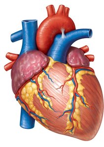
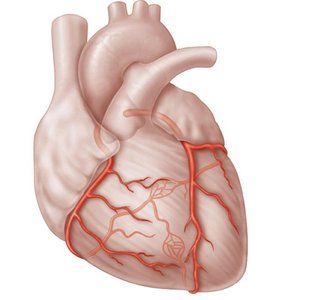
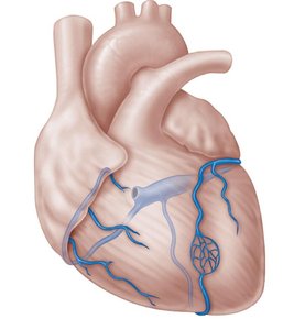
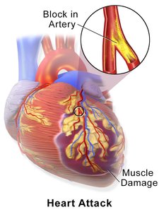
The heart muscle (myocardium) receives its own blood supply via the coronary arteries and is drained by cardiac veins that merge into the coronary sinus.
Coronary arteries: Branch from the aorta and supply oxygen-rich blood to the heart wall.
Cardiac veins: Drain deoxygenated blood from the myocardium into the coronary sinus, which empties into the right atrium.
Clinical Note: Blockage of coronary arteries can lead to angina pectoris (chest pain) or myocardial infarction (heart attack).
Microscopic Structure of Cardiac Muscle
Cardiac Muscle Cell Structure
Cardiac muscle cells are striated, short, branched, and interconnected by intercalated discs. Each cell typically has one central nucleus.
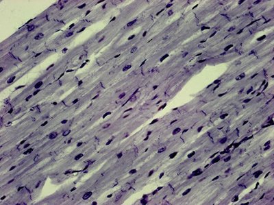
Intercalated discs: Specialized junctions containing gap junctions (for electrical coupling) and desmosomes (for mechanical strength).
Comparison with Skeletal Muscle
Both cardiac and skeletal muscle contract in response to action potentials and require calcium for contraction.
Cardiac muscle cells are mostly self-excitable (pacemaker cells), contract as a unit, and rely almost exclusively on aerobic respiration.
Electrical Activity and the Cardiac Conduction System
Pacemaker Cells and Action Potentials
The heart's intrinsic conduction system consists of pacemaker cells that generate and distribute electrical impulses, ensuring coordinated contraction.
Sinoatrial (SA) node: Pacemaker of the heart, initiates each heartbeat.
Atrioventricular (AV) node: Delays impulse, allowing atria to contract before ventricles.
AV bundle (Bundle of His): Only electrical connection between atria and ventricles.
Right and left bundle branches: Conduct impulses through interventricular septum.
Purkinje fibers: Distribute impulse through ventricular walls.
Action Potential Phases in Cardiac Cells
Pacemaker potential: Slow depolarization due to Na+ influx.
Depolarization: Ca2+ influx triggers action potential.
Repolarization: K+ efflux restores resting potential.
Electrocardiography (ECG)
Waves and Intervals
An ECG records the electrical activity of the heart. The main components are:
P wave: Atrial depolarization
QRS complex: Ventricular depolarization (and atrial repolarization)
T wave: Ventricular repolarization
Abnormal ECG patterns can indicate arrhythmias, heart blocks, or fibrillation.
Mechanical Events: The Cardiac Cycle
Phases of the Cardiac Cycle
The cardiac cycle consists of all events associated with blood flow through the heart during one heartbeat:
Ventricular filling: AV valves open, blood flows into ventricles.
Atrial contraction: Completes ventricular filling (end-diastolic volume, EDV).
Isovolumetric contraction: Ventricles contract, AV valves close, SL valves not yet open.
Ventricular ejection: SL valves open, blood ejected into aorta and pulmonary trunk.
Isovolumetric relaxation: Ventricles relax, SL valves close, blood remaining is end-systolic volume (ESV).
Heart sounds: "Lub-dup" sounds correspond to closure of AV valves (lub) and SL valves (dup).
Volume and Pressure Changes
Stroke volume (SV): Volume of blood pumped by each ventricle per beat.
EDV: Volume in ventricle at end of diastole (filling).
ESV: Volume in ventricle at end of systole (contraction).
Formula:
Regulation of Cardiac Output
Cardiac Output (CO)
Cardiac output is the amount of blood pumped by each ventricle in one minute.
Formula:
Typical resting CO: 5.25 L/min (HR = 75 bpm, SV = 70 ml/beat)
Factors Affecting Stroke Volume
Preload: Degree of stretch of cardiac muscle before contraction (influenced by venous return).
Contractility: Force of contraction at a given preload (increased by sympathetic stimulation).
Afterload: Pressure ventricles must overcome to eject blood (increased in hypertension).
Regulation of Heart Rate
Autonomic nervous system: Sympathetic stimulation increases HR and contractility; parasympathetic (vagus nerve) decreases HR.
Vagal tone: Parasympathetic dominance maintains resting HR at ~75 bpm.
Abnormal Heart Rates and Heart Failure
Tachycardia: Resting HR > 100 bpm; may lead to fibrillation.
Bradycardia: Resting HR < 60 bpm; may result from endurance training or cause inadequate circulation.
Heart failure: Inability of the heart to supply adequate blood flow; may result from coronary artery disease, hypertension, or myocardial infarction.
Summary Table: Key Cardiac Formulas
Parameter | Formula | Description |
|---|---|---|
Cardiac Output (CO) | Blood pumped per minute | |
Stroke Volume (SV) | Blood pumped per beat |
Additional info: This guide covers the structure, function, and physiology of the heart as outlined in a typical college-level anatomy and physiology course.
