 Back
BackThe Cardiovascular System: The Heart – Structure, Function, and Organization
Study Guide - Smart Notes
 Tailored notes based on your materials, expanded with key definitions, examples, and context.
The Cardiovascular System: The Heart
Introduction to the Heart
The heart is a muscular organ essential for maintaining the circulation of blood throughout the body. It ensures the delivery of oxygen and nutrients to tissues and the removal of metabolic wastes. On average, the heart beats about 100,000 times per day, pumping approximately 1,500,000 gallons of blood each year. The rate of blood flow can vary depending on individual activity levels.
Function: Maintains continuous blood flow to sustain life.
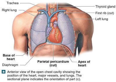
Location: Situated in the mediastinum of the thoracic cavity, between the lungs.
Size: Roughly the size of two clenched fists.
Overview of the Cardiovascular System
Circuits and Blood Vessels
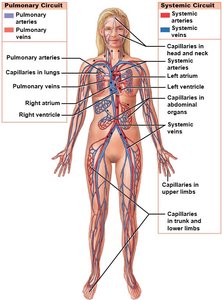
The heart pumps blood through two main circuits: the pulmonary circuit and the systemic circuit. Each circuit contains three types of blood vessels: arteries, veins, and capillaries.
Pulmonary Circuit: Carries deoxygenated blood to the lungs for oxygenation and returns oxygenated blood to the heart.
Systemic Circuit: Delivers oxygenated blood to the body and returns deoxygenated blood to the heart.
Arteries: Transport blood away from the heart.
Veins: Transport blood toward the heart.
Capillaries: Tiny vessels where gas and nutrient exchange occurs.
Pericardium and Heart Wall Structure
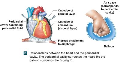
The Pericardium
The heart is enclosed by the pericardium, a double-layered sac that reduces friction and protects the heart. The pericardium consists of:
Fibrous Pericardium: Outer dense connective tissue layer.
Serous Pericardium: Inner membrane with parietal and visceral layers.
Pericardial Cavity: Space between the serous layers containing lubricating fluid.
Layers of the Heart Wall
The heart wall is composed of three layers:
Epicardium: Outer layer, also known as the visceral layer of the serous pericardium; made of simple squamous epithelium (mesothelium).
Myocardium: Thick, muscular middle layer containing cardiac muscle tissue, connective tissue, blood vessels, and nerves.
Endocardium: Inner layer of simple squamous epithelium (endothelium) continuous with the lining of blood vessels.
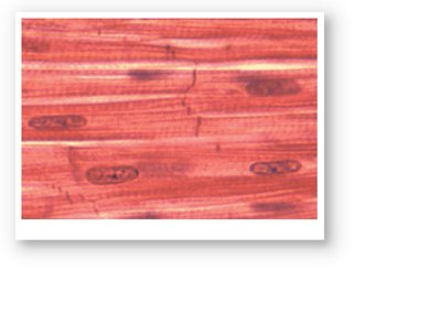
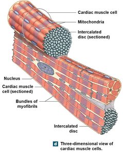
Cardiac Muscle Tissue and Intercalated Discs
Cardiac muscle cells are short, striated, and typically have a single nucleus. They are highly vascularized and rely on aerobic respiration. The cells are interconnected by specialized junctions called intercalated discs, which contain desmosomes and gap junctions.
Intercalated Discs: Strengthen connections between cells and allow rapid electrical communication via gap junctions.
Function: Enables the heart to contract as a coordinated unit.
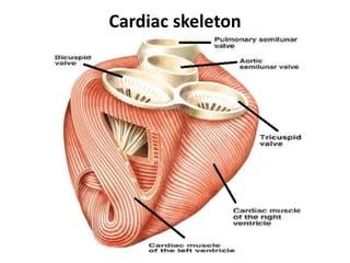
The Cardiac Skeleton
The cardiac skeleton is a framework of dense connective tissue that provides structural support, maintains heart shape, and prevents overstretching. It also electrically isolates the atria from the ventricles, ensuring proper timing of contractions.
Attachment: Anchors cardiac muscle fibers and heart valves.
Function: Distributes force of contraction and maintains chamber integrity.
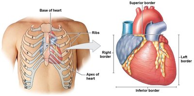
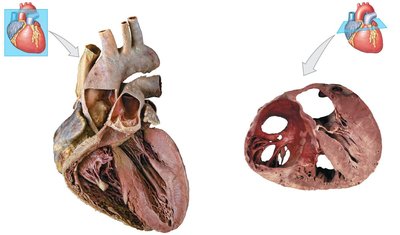
Orientation and Superficial Anatomy of the Heart
External Features and Landmarks
The heart is oriented with its base (superior border) at the top and apex (inferior tip) pointing left. Major external landmarks include:
Base: Where major vessels enter/exit, located posterior to the third costal cartilage.
Apex: Inferior tip, posterior to the fifth costal cartilage.
Sulci: Surface grooves filled with adipose tissue, marking chamber boundaries (coronary sulcus, anterior/posterior interventricular sulci).
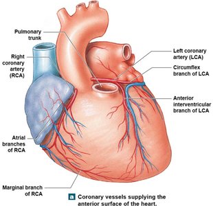
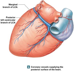
Coronary Blood Vessels
Coronary arteries supply oxygenated blood to the heart muscle, while coronary veins return deoxygenated blood to the right atrium via the coronary sinus.
Coronary Arteries: Branch from the aorta and run in the sulci.
Coronary Veins: Drain into the coronary sinus.
Internal Anatomy and Organization of the Heart
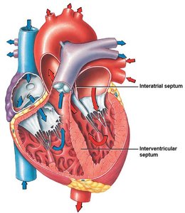
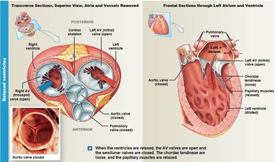
Chambers and Valves
The heart contains four chambers: two atria (receiving chambers) and two ventricles (pumping chambers). Valves ensure unidirectional blood flow:
Atrioventricular (AV) Valves: Tricuspid (right) and mitral/bicuspid (left) valves between atria and ventricles.
Semilunar Valves: Pulmonary (right ventricle to pulmonary artery) and aortic (left ventricle to aorta) valves.
Right Side of the Heart
The right atrium receives deoxygenated blood from the superior and inferior vena cava and the coronary sinus. Blood passes through the tricuspid valve into the right ventricle, which pumps it through the pulmonary valve into the pulmonary trunk and arteries.
Pectinate Muscles: Found in the right atrium, increase contraction strength.
Moderator Band: Unique to the right ventricle, helps coordinate contraction.
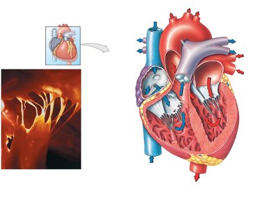
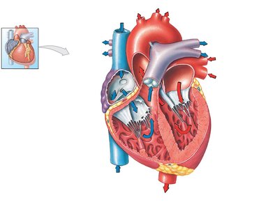
Left Side of the Heart
Oxygenated blood returns from the lungs via four pulmonary veins to the left atrium. It passes through the mitral valve into the left ventricle, which has the thickest walls to generate enough force to pump blood throughout the body. Blood exits through the aortic valve into the aorta.
Trabeculae Carneae: Muscular ridges in the ventricles, more prominent in the left ventricle.
Papillary Muscles: Two in the left ventricle, attached to the mitral valve via chordae tendineae.
Valve Function and Cardiac Cycle
Heart valves open and close in response to pressure changes during the cardiac cycle:
Diastole (Relaxation): AV valves open, semilunar valves closed; ventricles fill with blood.
Systole (Contraction): Ventricles contract, AV valves close to prevent backflow, semilunar valves open to allow blood ejection.
The Coordination of Cardiac Contractions
Conducting System
Cardiac contractions are coordinated by pacemaker cells, primarily located in the sinoatrial (SA) node. The electrical impulse travels from the SA node through the atria to the atrioventricular (AV) node, down the interventricular septum via the bundle of His, and then through Purkinje fibers to the ventricular myocardium.
SA Node: Main pacemaker, sets rhythm (60–100 bpm).
AV Node and Purkinje Fibers: Serve as backup pacemakers.
Autonomic Control of Heart Rate
The autonomic nervous system (ANS) modulates heart rate to maintain homeostasis. The cardiovascular control centers (CVCC) in the brainstem receive sensory input and adjust heart rate via sympathetic (increases rate) and parasympathetic (decreases rate) pathways.
Sympathetic Stimulation: Increases heart rate and force of contraction.
Parasympathetic Stimulation: Decreases heart rate.
Summary Table: Heart Chambers and Valves
Chamber | Receives Blood From | Pumps Blood To | Valve |
|---|---|---|---|
Right Atrium | Superior/Inferior Vena Cava, Coronary Sinus | Right Ventricle | Tricuspid (AV) Valve |
Right Ventricle | Right Atrium | Pulmonary Trunk | Pulmonary Semilunar Valve |
Left Atrium | Pulmonary Veins | Left Ventricle | Mitral (Bicuspid) Valve |
Left Ventricle | Left Atrium | Aorta | Aortic Semilunar Valve |
Key Terms and Definitions
Myocardium: Muscular middle layer of the heart wall.
Intercalated Discs: Specialized junctions connecting cardiac muscle cells.
Pacemaker Cells: Cells that generate rhythmic electrical impulses.
Cardiac Skeleton: Dense connective tissue framework supporting the heart.
Trabeculae Carneae: Muscular ridges in the ventricles.
Pectinate Muscles: Muscular ridges in the atria.
Example: Blood Flow Through the Heart
Deoxygenated blood enters the right atrium from the body (via vena cavae and coronary sinus).
Passes through the tricuspid valve into the right ventricle.
Pumped through the pulmonary valve into the pulmonary trunk and arteries to the lungs.
Oxygenated blood returns via pulmonary veins to the left atrium.
Passes through the mitral valve into the left ventricle.
Pumped through the aortic valve into the aorta and systemic circulation.
Additional info: The cardiac cycle consists of systole (contraction) and diastole (relaxation), and the heart's electrical system ensures coordinated contraction for efficient blood flow.
