 Back
The Cardiovascular System: The Heart – Structure, Function, and Circulation
Study Guide - Smart Notes
 Tailored notes based on your materials, expanded with key definitions, examples, and context.
Heart Anatomy and Orientation
Overview of Heart Structure and Function
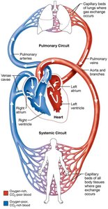
The heart is a muscular organ that functions as a double pump, circulating blood through two primary circuits: the pulmonary circuit (to and from the lungs) and the systemic circuit (to and from the body tissues). Its structure and location are optimized for efficient blood flow and protection within the thoracic cavity.
Right side: Receives oxygen-poor blood from the body and pumps it to the lungs for gas exchange (pulmonary circuit).
Left side: Receives oxygen-rich blood from the lungs and pumps it to the rest of the body (systemic circuit).
Chambers: Two atria (receiving chambers) and two ventricles (pumping chambers).
Size, Location, and Orientation of the Heart
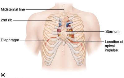
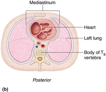
The heart is approximately the size of a fist and weighs less than 1 pound. It is located in the mediastinum, between the second rib and fifth intercostal space, resting on the superior surface of the diaphragm. About two-thirds of the heart lies to the left of the midsternal line, anterior to the vertebral column and posterior to the sternum. The base points toward the right shoulder, and the apex points toward the left hip.
Coverings and Layers of the Heart
Pericardium and Heart Wall Layers
The heart is enclosed in a double-walled sac called the pericardium:
Fibrous pericardium: Tough, superficial layer that protects, anchors, and prevents overfilling.
Serous pericardium: Deep, two-layered membrane (parietal and visceral layers) with a fluid-filled pericardial cavity that reduces friction.
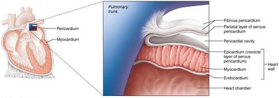
The heart wall consists of three layers:
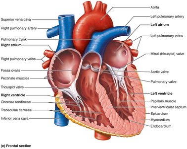
Epicardium: Outer layer, same as the visceral layer of the serous pericardium.
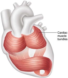
Myocardium: Middle, muscular layer responsible for contraction; contains spiral/circular bundles of cardiac muscle and the cardiac skeleton (connective tissue network).
Endocardium: Inner layer, continuous with the endothelial lining of blood vessels; lines chambers and covers valves.
Chambers, Septa, and Surface Features
Internal and External Anatomy
The heart has four chambers separated by septa and marked by surface grooves:
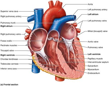
Interatrial septum: Separates the atria; contains the fossa ovalis (remnant of fetal foramen ovale).
Interventricular septum: Separates the ventricles.
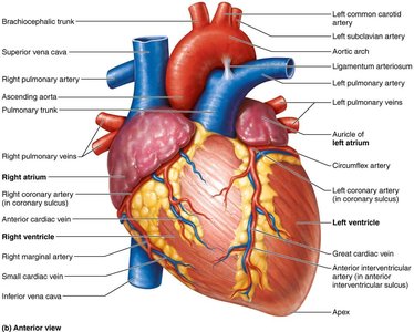
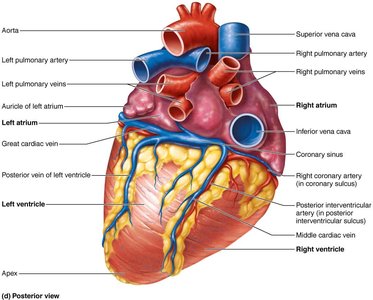
Coronary sulcus (atrioventricular groove): Encircles the junction between atria and ventricles.
Anterior and posterior interventricular sulci: Mark the position of the interventricular septum on the heart's surface.
Chamber Functions and Associated Vessels
Right atrium: Receives deoxygenated blood from the superior/inferior vena cava and coronary sinus.
Left atrium: Receives oxygenated blood from four pulmonary veins.
Right ventricle: Pumps blood into the pulmonary trunk (to lungs).
Left ventricle: Pumps blood into the aorta (to body); has the thickest wall due to high systemic pressure.
Heart Valves
Types and Functions of Valves
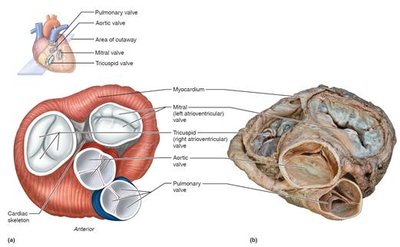
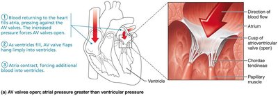
Heart valves ensure unidirectional blood flow and open/close in response to pressure changes:
Atrioventricular (AV) valves: Between atria and ventricles (tricuspid on right, mitral/bicuspid on left).
Semilunar (SL) valves: Between ventricles and major arteries (pulmonary SL valve on right, aortic SL valve on left).
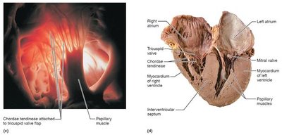
AV valves are supported by chordae tendineae anchored to papillary muscles, preventing valve prolapse during ventricular contraction.
Pathway of Blood Through the Heart
Blood Flow Sequence
Blood flows through the heart in a specific sequence, ensuring separation of oxygen-poor and oxygen-rich blood:
Body → Right atrium → Tricuspid valve → Right ventricle → Pulmonary semilunar valve → Pulmonary trunk → Pulmonary arteries → Lungs
Lungs → Pulmonary veins → Left atrium → Mitral valve → Left ventricle → Aortic semilunar valve → Aorta → Body
The right ventricle pumps blood to the low-pressure pulmonary circuit, while the left ventricle pumps to the high-pressure systemic circuit.
Coronary Circulation
Blood Supply to the Heart Muscle
The heart muscle (myocardium) receives its own blood supply via the coronary arteries, which branch from the base of the aorta. Major branches include:
Left coronary artery: Anterior interventricular artery, circumflex artery
Right coronary artery: Right marginal artery, posterior interventricular artery
Venous blood is collected by cardiac veins and returned to the right atrium via the coronary sinus.
Microscopic Anatomy of Cardiac Muscle
Cardiac Muscle Structure
Cardiac muscle cells are striated, short, branched, and interconnected by intercalated discs containing:
Desmosomes: Provide structural strength and prevent cell separation during contraction.
Gap junctions: Allow ions to pass, enabling electrical coupling and coordinated contraction (functional syncytium).
Cardiac muscle has abundant mitochondria (25–35% of cell volume), relies on aerobic metabolism, and has a less extensive sarcoplasmic reticulum than skeletal muscle.
Comparison: Cardiac vs. Skeletal Muscle
Feature | Cardiac Muscle | Skeletal Muscle |
|---|---|---|
Control of Action Potentials | Some cells are self-excitable (pacemaker cells) | Requires nervous system stimulation |
Contraction Pattern | All-or-none, functional syncytium | Motor units contract independently |
Calcium Source | Extracellular Ca2+ triggers SR release | Primarily from SR |
Refractory Period | Long, prevents tetanus | Short, tetanus possible |
Energy Use | Almost exclusively aerobic | Can use anaerobic fermentation |
Glossary of Key Terms
Pericardium: Double-walled sac enclosing the heart
Myocardium: Muscular, contractile middle layer of the heart wall
Endocardium: Inner lining of the heart chambers and valves
Chordae tendineae: Collagen cords anchoring AV valve flaps to papillary muscles
Trabeculae carneae: Irregular muscle ridges on ventricular walls
Fossa ovalis: Remnant of the fetal foramen ovale in the interatrial septum
Coronary sinus: Large vein collecting blood from the myocardium and delivering it to the right atrium
