 Back
The Cardiovascular System: The Heart – Structure, Function, and Physiology
Study Guide - Smart Notes
 Tailored notes based on your materials, expanded with key definitions, examples, and context.
Heart Anatomy
Location of the Heart
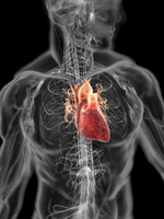
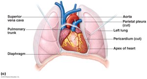
The heart is a muscular organ located in the mediastinum, resting on the superior surface of the diaphragm. Approximately two-thirds of the heart lies to the left of the midsternal line, positioned anterior to the vertebral column and posterior to the sternum. It is roughly the size of a fist and is enclosed within the pericardium, a double-walled sac that provides protection and anchorage.
Pericardium
The pericardium consists of two main layers:
Superficial fibrous pericardium: Protects, anchors, and prevents overfilling of the heart.
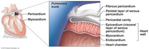
Deep serous pericardium: Composed of two layers separated by a fluid-filled pericardial cavity, which reduces friction during heart activity.
Parietal layer: Lines the internal surface of the fibrous pericardium.
Visceral layer (epicardium): Covers the external surface of the heart.
Layers of the Heart Wall
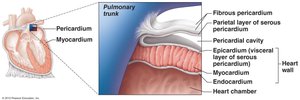
The heart wall is composed of three layers:
Epicardium: The visceral layer of the serous pericardium.
Myocardium: Consists of spiral bundles of cardiac muscle cells and the fibrous skeleton of the heart, which anchors muscle fibers, supports vessels and valves, and limits the spread of action potentials.
Endocardium: A thin endothelial layer lining the heart chambers and continuous with blood vessel endothelium.
Chambers of the Heart
Atria: The Receiving Chambers
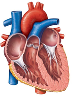
The heart contains four chambers: two atria and two ventricles. The atria are separated by the interatrial septum and are externally marked by auricles, which increase atrial volume. The right atrium receives blood from the superior and inferior vena cava and the coronary sinus, while the left atrium receives blood from the right and left pulmonary veins.
Ventricles: The Discharging Chambers
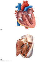
The ventricles are separated by the interventricular septum and are externally marked by anterior and posterior interventricular sulci. The right ventricle pumps blood into the pulmonary trunk (to the lungs), and the left ventricle pumps blood into the aorta (to the body). The walls of the ventricles are ridged by trabeculae carneae, and papillary muscles project into the ventricular cavities.
Pathway of Blood Through the Heart
Pulmonary and Systemic Circuits
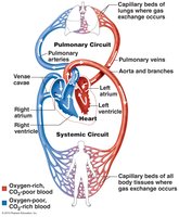
The heart functions as two side-by-side pumps:
Right side: Pumps blood through the pulmonary circuit (to and from the lungs).
Left side: Pumps blood through the systemic circuit (to and from body tissues).
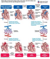
Blood pathway: Right atrium → tricuspid valve → right ventricle → pulmonary semilunar valve → pulmonary trunk → pulmonary arteries → lungs → pulmonary veins → left atrium → bicuspid (mitral) valve → left ventricle → aortic semilunar valve → aorta → systemic circulation.
Coronary Circulation
Arterial and Venous Supply
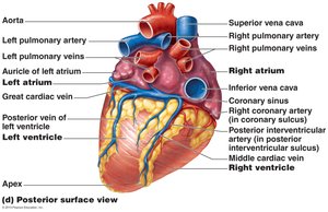
Coronary circulation provides the functional blood supply to the heart muscle itself. The right and left coronary arteries branch into several arteries, including the marginal, posterior interventricular, circumflex, and anterior interventricular arteries. Venous blood is collected by the small, anterior, and great cardiac veins, which join to form the coronary sinus.
Homeostatic Imbalances
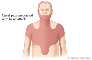
Angina pectoris: Thoracic pain due to transient deficiency in blood delivery to the myocardium; cells are weakened.
Myocardial infarction (heart attack): Prolonged coronary blockage leads to cell death, which is replaced by noncontractile scar tissue.
Heart Valves
Atrioventricular (AV) Valves
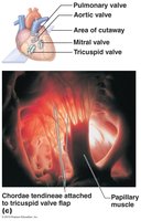
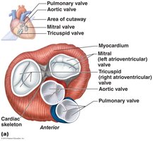
AV valves prevent backflow into the atria when the ventricles contract. The right AV valve is the tricuspid valve, and the left is the mitral (bicuspid) valve. Chordae tendineae anchor the valve cusps to papillary muscles.
Semilunar (SL) Valves
SL valves prevent backflow into the ventricles when the ventricles relax. The aortic semilunar valve is located at the left ventricle, and the pulmonary semilunar valve is at the right ventricle.
Microscopic Anatomy of Cardiac Muscle
Cardiac Muscle Structure
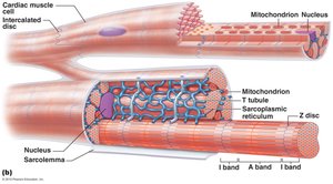
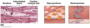
Cardiac muscle cells are striated, short, branched, and interconnected. They contain numerous mitochondria and a connective tissue matrix (endomysium) that connects to the fibrous skeleton. Intercalated discs, containing desmosomes and gap junctions, anchor cells and allow for electrical coupling, enabling the heart to function as a syncytium.
Cardiac Muscle Contraction
Mechanism of Contraction
Cardiac muscle contraction is initiated by depolarization, which opens voltage-gated fast Na+ channels, reversing the membrane potential. The depolarization wave in T tubules causes the sarcoplasmic reticulum to release Ca2+, and slow Ca2+ channels in the sarcolemma prolong the depolarization phase (plateau). Ca2+ influx triggers further Ca2+ release from the SR, leading to contraction. Repolarization occurs as Ca2+ channels close and K+ channels open.
Heart Physiology: Electrical Events
Intrinsic Cardiac Conduction System
The intrinsic conduction system consists of autorhythmic cells that initiate and distribute impulses, coordinating heart depolarization and contraction. The sequence of excitation is as follows:
Sinoatrial (SA) node: Pacemaker, generates impulses (~75/min).
Atrioventricular (AV) node: Delays impulses (~0.1 s), allows atria to contract before ventricles.
AV bundle (bundle of His): Only electrical connection between atria and ventricles.
Right and left bundle branches: Carry impulses through the interventricular septum.
Purkinje fibers: Complete the pathway into the apex and ventricular walls.
Homeostatic Imbalances
Arrhythmias: Irregular heart rhythms.
Fibrillation: Rapid, irregular contractions; ineffective for pumping blood.
Ectopic focus: Abnormal pacemaker activity.
Heart block: Impaired conduction between atria and ventricles.
Extrinsic Innervation of the Heart
The autonomic nervous system (ANS) modifies heart rate and force. The cardioacceleratory center (sympathetic) increases heart rate and contractility, while the cardioinhibitory center (parasympathetic, via the vagus nerve) decreases heart rate.
Electrocardiography (ECG/EKG)
An ECG records the electrical activity of the heart. Key waves include:
P wave: Depolarization of the SA node (atrial depolarization).
QRS complex: Ventricular depolarization.
T wave: Ventricular repolarization.
Heart Sounds
Two main heart sounds are produced by valve closure:
First sound (lub): AV valves close (beginning of systole).
Second sound (dup): SL valves close (beginning of diastole).
Heart murmurs are abnormal sounds, often indicating valve problems.
Mechanical Events: The Cardiac Cycle
Phases of the Cardiac Cycle
Ventricular filling: AV valves open, blood flows into ventricles; atrial systole delivers remaining blood.
Ventricular systole: Ventricles contract, AV valves close, SL valves open for ejection phase.
Isovolumetric relaxation: Ventricles relax, SL valves close, brief rise in aortic pressure (dicrotic notch).
End diastolic volume (EDV): Volume in ventricles at end of diastole. End systolic volume (ESV): Volume remaining after contraction.
Cardiac Output (CO)
Cardiac output is the volume of blood pumped by each ventricle per minute:
Where:
HR: Heart rate (beats per minute)
SV: Stroke volume (volume pumped per beat)
Stroke volume is calculated as:
Cardiac reserve is the difference between resting and maximal CO.
Regulation of Stroke Volume
Three main factors affect stroke volume:
Preload: Degree of stretch of cardiac muscle before contraction (Frank-Starling law).
Contractility: Contractile strength at a given muscle length, independent of preload.
Afterload: Pressure that must be overcome to eject blood (mainly arterial pressure).
Regulation of Heart Rate
Heart rate is regulated by:
Autonomic nervous system: Sympathetic stimulation increases HR (positive chronotropic effect); parasympathetic stimulation decreases HR (negative chronotropic effect).
Other factors: Age, gender, exercise, and body temperature.
Homeostatic Imbalances and Heart Failure
Tachycardia: Abnormally fast heart rate (>100 bpm).
Bradycardia: Abnormally slow heart rate (<60 bpm).
Congestive heart failure (CHF): Progressive condition where CO is too low to meet tissue needs, caused by coronary atherosclerosis, high blood pressure, multiple infarcts, or dilated cardiomyopathy.
Age-Related and Developmental Aspects
Sclerosis and thickening of valve flaps
Decline in cardiac reserve
Fibrosis of cardiac muscle
Atherosclerosis
Congenital heart defects: Mixing of systemic and pulmonary blood, narrowed valves or vessels
