 Back
The Cardiovascular System: The Heart – Structure, Function, and Physiology
Study Guide - Smart Notes
 Tailored notes based on your materials, expanded with key definitions, examples, and context.
The Cardiovascular System: The Heart
Overview
The heart is a muscular organ responsible for pumping blood throughout the body via the pulmonary and systemic circuits. It is essential for delivering oxygen and nutrients to tissues and removing waste products. This chapter covers the anatomy, physiology, and development of the heart, as well as common clinical conditions.
Heart Anatomy
Location and Size
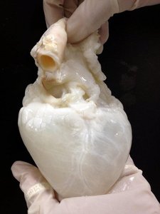
The heart is approximately the size of a fist and weighs less than 1 pound.
It is located in the mediastinum, on the superior surface of the diaphragm, left of the midsternal line, anterior to the vertebral column, and posterior to the sternum.
The base is at the top, and the apex is at the bottom, where the apical impulse can be felt.
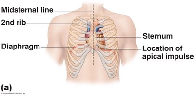
Coverings of the Heart: The Pericardium
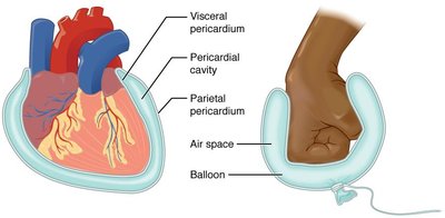
Pericardium: A double-walled sac that encloses the heart.
Consists of:
Fibrous pericardium: Superficial, protects, anchors, and prevents overfilling.
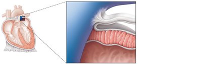
Serous pericardium: Deep, two-layered (parietal and visceral/epicardium) with a fluid-filled pericardial cavity that reduces friction.
Pericardial Disorders
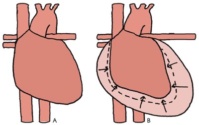
Pericarditis: Inflammation of the pericardium, causing friction rub and possibly leading to cardiac tamponade (compression of the heart by excess fluid).
Treatment involves draining excess fluid.
Layers of the Heart Wall
Epicardium: Visceral layer of serous pericardium, may contain fat in older individuals.
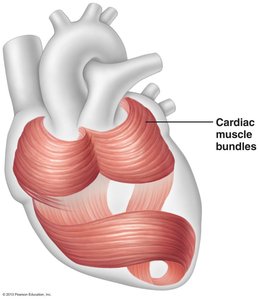
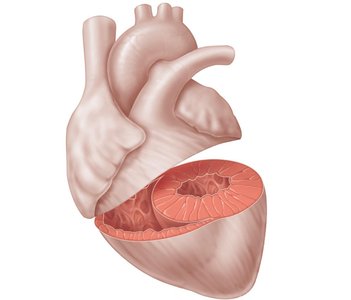
Myocardium: Cardiac muscle layer, responsible for contraction; contains cardiac muscle bundles and the cardiac skeleton (fibrous tissue network).
Endocardium: Lines the heart chambers and continues into blood vessels.
Chambers of the Heart
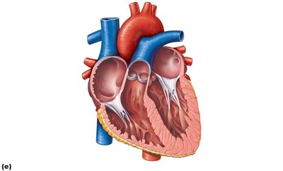
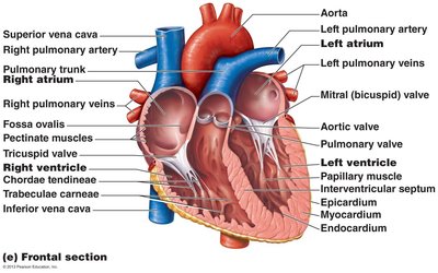
Four chambers: two superior atria (right and left) and two inferior ventricles (right and left).
Interatrial septum separates atria; interventricular septum separates ventricles.
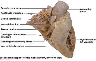
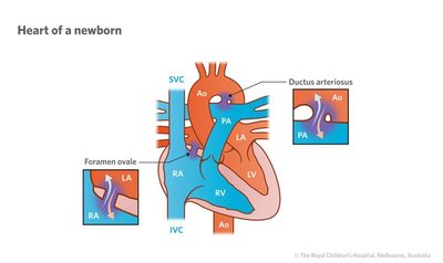
Fossa ovalis is a remnant of the fetal foramen ovale.
Atria: The Receiving Chambers
Small, thin-walled chambers that receive blood returning to the heart.
Right atrium receives blood from the systemic circuit (superior/inferior vena cava, coronary sinus).
Left atrium receives blood from the pulmonary circuit (four pulmonary veins).
Auricles increase atrial volume; pectinate muscles are present in the right atrium and auricles.
Ventricles: The Discharging Chambers
Thick-walled chambers that pump blood out of the heart.
Right ventricle pumps blood into the pulmonary trunk (to the lungs).
Left ventricle pumps blood into the aorta (to the body).
Trabeculae carneae and papillary muscles are present in the ventricles.
Heart Valves
Types and Functions
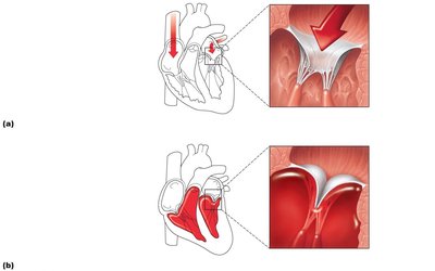
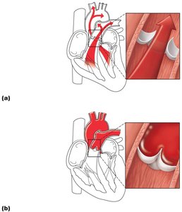
Valves ensure unidirectional blood flow and open/close in response to pressure changes.
Atrioventricular (AV) valves:
Tricuspid valve (right AV valve)
Mitral (bicuspid) valve (left AV valve)
Prevent backflow into atria during ventricular contraction; chordae tendineae anchor cusps to papillary muscles.
Semilunar (SL) valves:
Aortic semilunar valve
Pulmonary semilunar valve
Prevent backflow into ventricles during relaxation.
Valve Disorders
Incompetent valve: Blood backflows, requiring the heart to repump the same blood.
Valvular stenosis: Stiff valve flaps constrict the opening, increasing the heart's workload.
Valves may be replaced with mechanical, animal, or cadaver valves if severely damaged.
Pathway of Blood Through the Heart
Pulmonary and Systemic Circuits
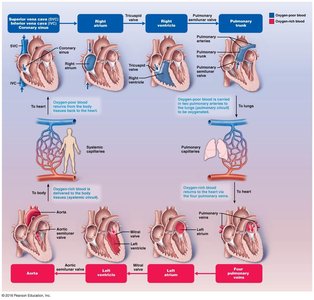
Pulmonary circuit: Right atrium → tricuspid valve → right ventricle → pulmonary semilunar valve → pulmonary trunk → pulmonary arteries → lungs → pulmonary veins → left atrium.
Systemic circuit: Left atrium → mitral valve → left ventricle → aortic semilunar valve → aorta → systemic circulation.
Equal volumes of blood are pumped to both circuits, but the systemic circuit is longer and higher pressure.
Coronary Circulation
Supplying the Heart Muscle
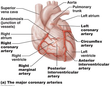
Coronary arteries arise from the base of the aorta and supply oxygenated blood to the heart muscle.
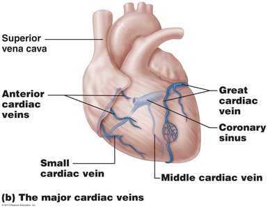
Cardiac veins drain deoxygenated blood from the myocardium into the coronary sinus, which empties into the right atrium.
Microscopic Anatomy of Cardiac Muscle
Structure and Function
Cardiac muscle cells are striated, branched, and interconnected by intercalated discs.
Desmosomes prevent cell separation during contraction; gap junctions allow ions to pass, enabling the heart to function as a syncytium (single coordinated unit).
Cardiac muscle has more mitochondria than skeletal muscle, making it highly resistant to fatigue but dependent on oxygen.
Unique troponin isoforms allow early detection of heart muscle damage.
Heart Physiology: Setting the Basic Rhythm
Intrinsic Cardiac Conduction System
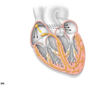
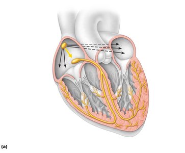
The heart can depolarize and contract without nervous system stimulation due to autorhythmic (pacemaker) cells.
The sequence of excitation:
Sinoatrial (SA) node (pacemaker) generates impulses.
Impulses pause at the atrioventricular (AV) node.
AV bundle connects atria to ventricles.
Bundle branches conduct impulses through the interventricular septum.
Subendocardial conducting network (Purkinje fibers) depolarizes ventricular cells.
Electrocardiography (ECG/EKG)
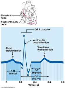
An ECG records the electrical activity of the heart.
Key waves:
P wave: Atrial depolarization
QRS complex: Ventricular depolarization and atrial repolarization
T wave: Ventricular repolarization
Mechanical Events: The Cardiac Cycle
Phases of the Cardiac Cycle
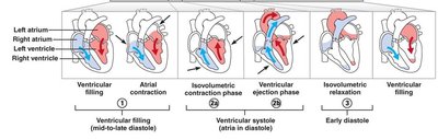
Ventricular filling (mid-to-late diastole): AV valves open, blood flows into ventricles, atrial systole delivers remaining blood.
Ventricular systole: Atria relax, ventricles contract, AV valves close, SL valves open for ejection phase.
Isovolumetric relaxation (early diastole): Ventricles relax, SL valves close, AV valves reopen when atrial pressure exceeds ventricular pressure.
Cardiac Output (CO)
CO is the volume of blood pumped by each ventricle per minute.
Formula:
Normal CO is about 5.25 L/min at rest.
Cardiac reserve is the difference between resting and maximal CO.
Regulation of Heart Rate
Neural and Chemical Regulation
Autonomic Nervous System (ANS):
Sympathetic division increases heart rate (norepinephrine, epinephrine).
Parasympathetic division decreases heart rate (vagal tone).
Chemical regulation:
Hormones (epinephrine, thyroxine) increase heart rate.
Ions (Ca2+, K+) must be balanced for normal function.
Other factors: age, gender, exercise, and body temperature.
Homeostatic Imbalances
Arrhythmias: Irregular heart rhythms.
Fibrillation: Rapid, uncoordinated contractions; requires defibrillation.
Tachycardia: Abnormally fast heart rate (>100 bpm).
Bradycardia: Abnormally slow heart rate (<60 bpm).
Congestive heart failure (CHF): Inadequate cardiac output due to weakened myocardium.
Pulmonary congestion: Left heart failure; blood backs up in lungs.
Peripheral congestion: Right heart failure; blood pools in body organs.
Diagnosis of Heart Disorders
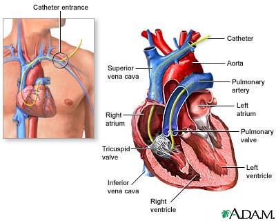
Catheterization: Inserting a catheter into the heart via blood vessels for diagnostic purposes.
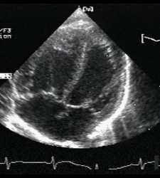
Echocardiography: Using ultrasound to visualize heart structures and function.
Developmental Aspects of the Heart
Embryonic and Fetal Heart
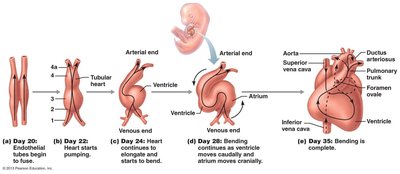
The heart begins as two tubes that fuse and form four chambers.
Heartbeat is detectable by 6 weeks of gestation.
Fetal heart structures (foramen ovale, ductus arteriosus) bypass pulmonary circulation and close after birth.
Congenital Heart Defects
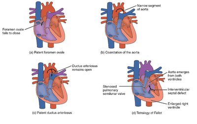
Most common birth defects; may require surgical correction.
Examples: patent foramen ovale, patent ductus arteriosus, coarctation of the aorta, tetralogy of Fallot.
Age-Related Changes
Sclerosis and thickening of valve flaps (heart murmurs more common in elderly).
Decline in cardiac reserve and maximum heart rate.
Fibrosis of cardiac muscle (replaced by scar tissue).
Atherosclerosis (accelerated by inactivity, smoking, stress, and poor diet).
