 Back
BackThe Cardiovascular System: The Heart – Study Notes
Study Guide - Smart Notes
 Tailored notes based on your materials, expanded with key definitions, examples, and context.
Introduction to the Cardiovascular System
Overview of Circuits
The cardiovascular system is responsible for transporting blood throughout the body, delivering oxygen and nutrients, and removing waste products. It consists of two main circuits:
Pulmonary Circuit: Carries blood to and from the gas exchange surfaces of the lungs. Blood is propelled by the contraction of the right ventricle.
Systemic Circuit: Carries blood to and from the rest of the body. Blood is propelled by the contraction of the left ventricle.
Blood alternates between the pulmonary and systemic circuits.
Types of Blood Vessels
Arteries: Carry blood away from the heart (usually oxygenated; color-coded red in textbooks).
Veins: Carry blood toward the heart (usually deoxygenated; color-coded blue in textbooks).
Capillaries: Smallest blood vessels; networks between arteries and veins. Enable exchange of gases, nutrients, and wastes between blood and tissues.
Structure and Anatomy of the Heart
Chambers of the Heart
The heart is a four-chambered organ that pumps oxygen-poor blood to the lungs and oxygen-rich blood to the body. The chambers are:
Right Atrium: Collects blood from the systemic circuit.
Right Ventricle: Pumps blood to the pulmonary circuit.
Left Atrium: Collects blood from the pulmonary circuit.
Left Ventricle: Pumps blood to the systemic circuit; has the thickest muscle layer and is the most powerful chamber.
Surface Anatomy and Pericardium
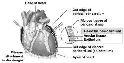
The heart is located in the mediastinum, between the two pleural cavities. It is surrounded by the pericardial sac and has a pointed apex and a broad base.
Pericardium: Double-layered lining of the pericardial cavity.
Parietal Pericardium: Outer layer, forms the inner layer of the pericardial sac.
Visceral Pericardium (Epicardium): Inner layer, covers the surface of the heart.
Pericardial Cavity: Space between parietal and visceral layers, contains pericardial fluid for lubrication.
Pericardial Sac: Fibrous tissue that surrounds and stabilizes the heart.
Heart Wall Layers
Epicardium: Outer layer (visceral pericardium).
Myocardium: Middle, muscular layer; responsible for contraction. Contains concentric layers of cardiac muscle tissue.
Endocardium: Inner layer; simple squamous epithelium lining the inner surfaces and heart valves.
Cardiac Muscle Tissue
Composed of cardiac muscle cells interconnected by intercalated discs.
Intercalated discs are secured by desmosomes and linked by gap junctions, allowing rapid propagation of action potentials and coordinated contractions.
Internal Anatomy and Organization
Septa and Valves
Interatrial Septum: Separates the two atria.
Interventricular Septum: Separates the two ventricles.
Atrioventricular (AV) Valves: Permit blood flow from atria to ventricles only.
Tricuspid Valve: Right AV valve; three cusps.
Bicuspid (Mitral) Valve: Left AV valve; two cusps.
Blood Flow Through the Heart
Right atrium receives blood from the body via the superior and inferior vena cava and the coronary sinus.
Right ventricle pumps blood to the lungs via the pulmonary trunk.
Left atrium receives blood from the lungs via four pulmonary veins.
Left ventricle pumps blood to the body via the aorta.
Heart Valves
AV Valves: Prevent backflow into atria during ventricular contraction; chordae tendineae and papillary muscles prevent valve inversion.
Semilunar Valves: Pulmonary and aortic; prevent backflow into ventricles during relaxation; have three cusps and no muscular support.
Coronary Circulation
Coronary Arteries: Originate at aortic sinuses; supply blood to the myocardium.
Cardiac Veins: Return blood to the right atrium via the coronary sinus.
Arterial Anastomoses: Interconnect anterior and posterior interventricular arteries, stabilizing blood supply to cardiac muscle.
The Conducting System
Cardiac Muscle Cell Types
Contractile Cells: Produce contractions that propel blood; action potential phases include rapid depolarization, plateau, and repolarization.
Conducting System: Specialized cells that initiate and distribute electrical impulses, ensuring coordinated contraction (automaticity).
Key Structures
Sinoatrial (SA) Node: Wall of right atrium; primary pacemaker (70–80 action potentials/min).
Atrioventricular (AV) Node: Junction between atria and ventricles; secondary pacemaker (40–60 action potentials/min).
Conducting Cells: Internodal pathways (atria), AV bundle and bundle branches (ventricles).
Electrocardiogram (ECG)
Records electrical events in the heart using electrodes.
P wave: Atrial depolarization.
QRS complex: Ventricular depolarization (systole).
T wave: Ventricular repolarization (diastole).
The Cardiac Cycle
Phases of the Cardiac Cycle
Systole: Contraction phase; blood pressure rises.
Diastole: Relaxation phase; blood pressure falls.
Blood flows from high to low pressure, directed by four one-way valves.
Heart Sounds
S1 ("lubb"): AV valves closing.
S2 ("dupp"): Semilunar valves closing.
S3, S4: Soft sounds (ventricular filling, atrial contraction).
Cardiodynamics
Cardiac Output
Cardiac output (CO) is the amount of blood pumped by the heart per minute. It is calculated as:
CO: Cardiac output (mL/min)
HR: Heart rate (beats/min)
SV: Stroke volume (mL/beat)
Factors Affecting Cardiac Output
Heart rate is adjusted by the autonomic nervous system and hormones.
Stroke volume is adjusted by changing end diastolic volume (EDV) or end systolic volume (ESV).
Blood Volume Reflexes
Atrial Reflex (Bainbridge Reflex): Adjusts heart rate in response to venous return; increased venous return stimulates stretch receptors in the right atrium, increasing heart rate via sympathetic activity.
Autonomic Innervation
Sympathetic Stimulation: Increases heart rate and force of contraction (norepinephrine, epinephrine).
Parasympathetic Stimulation: Decreases heart rate and force of contraction (acetylcholine via vagus nerve).
Cardiac Centers: Located in the medulla oblongata; cardioacceleratory (sympathetic) and cardioinhibitory (parasympathetic) centers.
Hormonal Effects
Epinephrine and norepinephrine increase heart rate.
Thyroid hormones influence heart rate and metabolism.
Pharmaceuticals can mimic or block these effects (e.g., beta blockers, calcium channel blockers).
Key Terms and Definitions
Atrium: Upper chamber of the heart that receives blood.
Myocardium: Muscular middle layer of the heart wall.
Myocardial Infarction (MI): Heart attack; death of heart muscle tissue due to lack of blood supply.
Diastole: Relaxation phase of the cardiac cycle.
Pericardium: Double-layered sac surrounding the heart.
Mitral (Left AV) Valve: Valve between left atrium and left ventricle.
Systole: Contraction phase of the cardiac cycle.
Bradycardia: Abnormally slow heart rate.
Tricuspid (Right AV) Valve: Valve between right atrium and right ventricle.
SA Node: Sinoatrial node; primary pacemaker of the heart.
Tachycardia: Abnormally fast heart rate.
Pulmonary Semilunar Valve: Valve between right ventricle and pulmonary trunk.
AV Node: Atrioventricular node; secondary pacemaker.
Purkinje Fibers: Conducting fibers that distribute electrical impulses through the ventricles.
Aortic Semilunar Valve: Valve between left ventricle and aorta.
Ventricle: Lower chamber of the heart that pumps blood out.
Coronary Arteries: Arteries supplying blood to the heart muscle.
