 Back
BackThe Cell: Structure, Function, and Membrane Transport
Study Guide - Smart Notes
 Tailored notes based on your materials, expanded with key definitions, examples, and context.
The Cell: Structure and Function
Cell Theory and Diversity
The cell is the fundamental structural and functional unit of all living organisms. The cell theory states that:
All living things are composed of cells.
Cells are the basic units of structure and function in living things.
All cells arise from pre-existing cells.
Cell function is dictated by cell shape and subcellular structures.
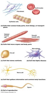
Human bodies contain over 200 different types of cells, each specialized in size, shape, and function.
Basic Parts of a Human Cell
Plasma membrane: Flexible outer boundary that separates the cell from its environment.
Cytoplasm: Intracellular fluid containing organelles.
Nucleus: Control center containing genetic material (DNA).
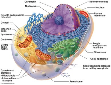
Cell Organelles and Their Functions
Organelle | Structure | Function |
|---|---|---|
Mitochondria | Double-membrane structure with cristae | Site of ATP synthesis; powerhouse of the cell |
Ribosomes | Dense particles of rRNA and protein | Site of protein synthesis |
Rough ER | Membranous system with ribosomes | Protein synthesis and transport |
Smooth ER | Membranous system without ribosomes | Lipid and steroid synthesis, detoxification |
Golgi apparatus | Stack of flattened membranes | Packages, modifies, and segregates proteins |
Lysosomes | Membranous sacs with acid hydrolases | Sites of intracellular digestion |
Peroxisomes | Membranous sacs of oxidase enzymes | Detoxify harmful substances |
The Plasma Membrane
Structure of the Plasma Membrane
The plasma membrane is a dynamic, selectively permeable barrier that separates the intracellular fluid from the extracellular environment. It is primarily composed of a phospholipid bilayer with embedded proteins, cholesterol, and carbohydrates.
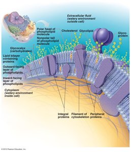
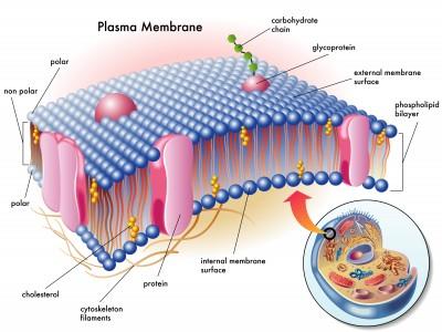
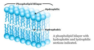
Phospholipid Bilayer
Phospholipids: Have hydrophilic (water-loving) heads and hydrophobic (water-fearing) tails, forming a bilayer that self-assembles and reseals if torn.
Cholesterol: Stabilizes the membrane and decreases the mobility of phospholipids.
Glycolipids: Lipids with attached sugars, important for cell recognition (e.g., blood types).
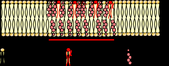
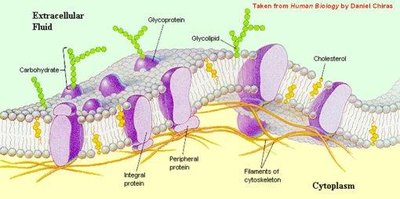
Membrane Proteins
Integral proteins: Span the membrane and are involved in transport and cell communication.
Peripheral proteins: Loosely attached to the membrane surface, involved in signaling and maintaining cell shape.
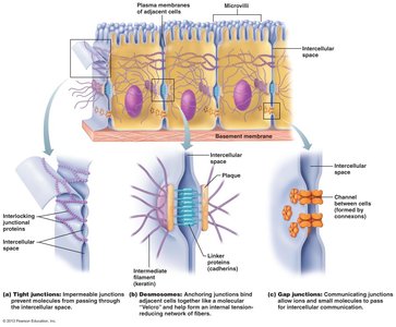
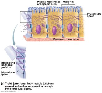
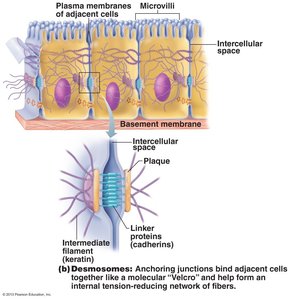
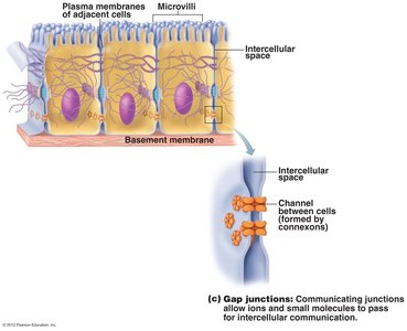
Cell Junctions
Types of Cell Junctions
Cells may exist as free entities or be bound together to form tissues. There are three main types of cell junctions:
Tight junctions: Impermeable junctions that prevent molecules from passing between cells (important in the digestive tract).
Desmosomes: Anchoring junctions that bind cells together like molecular "Velcro" (found in skin and heart muscle).
Gap junctions: Communicating junctions that allow ions and small molecules to pass for intercellular communication (important in cardiac and smooth muscle).
Membrane Transport
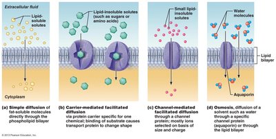
Passive Transport
Passive transport does not require cellular energy (ATP). Substances move down their concentration gradients.
Simple diffusion: Nonpolar and lipid-soluble substances diffuse directly through the lipid bilayer.
Facilitated diffusion: Transport of molecules via protein carriers or channels (e.g., glucose, ions).
Osmosis: Diffusion of water through a selectively permeable membrane.
Filtration: Movement of water and solutes across a membrane due to hydrostatic pressure (mainly in capillaries).
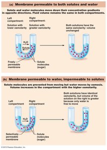
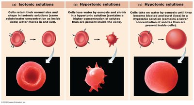
Tonicity
Isotonic solution: Same solute concentration as the cell; no net water movement.
Hypertonic solution: Higher solute concentration outside the cell; water leaves the cell, causing it to shrink.
Hypotonic solution: Lower solute concentration outside the cell; water enters the cell, causing it to swell and possibly burst.
Active Transport
Active transport requires energy (ATP) to move substances against their concentration gradients.
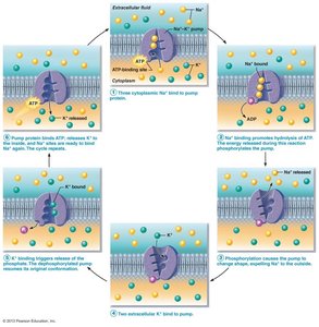
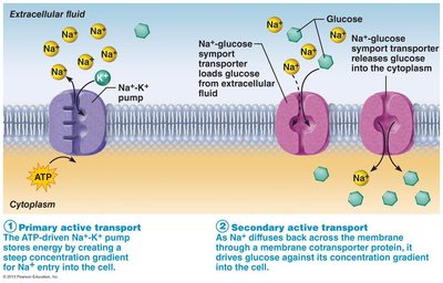
Primary active transport: Direct use of ATP (e.g., sodium-potassium pump).
Secondary active transport: Indirect use of ATP; uses energy stored in ionic gradients created by primary active transport.
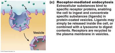
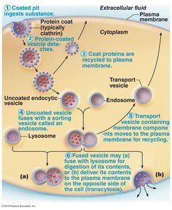
Vesicular transport: Movement of large particles and macromolecules via vesicles (endocytosis, exocytosis, transcytosis).
Vesicular Transport
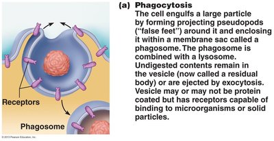
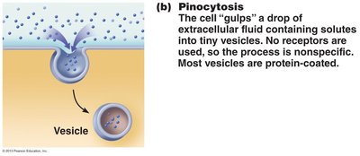
Endocytosis: Transport into the cell (phagocytosis, pinocytosis, receptor-mediated endocytosis).
Exocytosis: Transport out of the cell (e.g., hormone secretion, neurotransmitter release).
Transcytosis: Transport into, across, and out of the cell.
Vesicular trafficking: Transport from one area or organelle to another within the cell.
The Cell Cycle and Mitosis
Overview of the Cell Cycle
The cell cycle describes the series of events from cell formation to cell division. It consists of interphase (cell growth and DNA replication) and the mitotic phase (cell division).
Interphase: G1 (growth), S (DNA synthesis), G2 (preparation for division).
Mitotic phase: Mitosis (nuclear division) and cytokinesis (cytoplasmic division).
Phases of Mitosis
Prophase: Chromosomes condense, spindle forms, nuclear envelope breaks down.
Metaphase: Chromosomes align at the cell's equator.
Anaphase: Sister chromatids separate and move to opposite poles.
Telophase: Chromosomes decondense, nuclear envelope reforms.
Cytokinesis: Cytoplasm divides, forming two daughter cells.
DNA, RNA, and Protein Synthesis
Genetic Information Flow
DNA: Contains genes, which are instructions for synthesizing proteins.
RNA: Three types—mRNA (messenger), tRNA (transfer), rRNA (ribosomal).
Transcription: Synthesis of mRNA from DNA template in the nucleus.
Translation: Synthesis of polypeptides at the ribosome using mRNA code.
Key Steps in Protein Synthesis
Transcription: DNA → mRNA (in the nucleus)
RNA processing: Pre-mRNA is spliced and modified
Translation: mRNA → Protein (at the ribosome in the cytoplasm)
Summary Table: Types of RNA and Their Functions
Type of RNA | Function |
|---|---|
mRNA (messenger RNA) | Carries genetic code from DNA to ribosome |
tRNA (transfer RNA) | Brings amino acids to the ribosome during translation |
rRNA (ribosomal RNA) | Forms the core of the ribosome's structure and catalyzes protein synthesis |
Developmental Aspects and Cell Aging
Cell differentiation: Cells become specialized in structure and function during development.
Apoptosis: Programmed cell death, essential for development and tissue homeostasis.
Cell aging: Theories include wear and tear, mitochondrial dysfunction, immune system decline, and genetic programming (telomere shortening).
Key Study Points
Understand the structure and function of the plasma membrane, including membrane proteins and cell junctions.
Be able to explain membrane transport mechanisms (passive and active).
Know the phases of the cell cycle and mitosis.
Review the flow of genetic information from DNA to RNA to protein.
Animal cells do not have cell walls or chloroplasts.
