 Back
BackThe Cellular and Tissue Levels of Organization: Study Notes for Human Anatomy & Physiology I
Study Guide - Smart Notes
 Tailored notes based on your materials, expanded with key definitions, examples, and context.
The Cellular Level of Organization
Overview of Cell Types
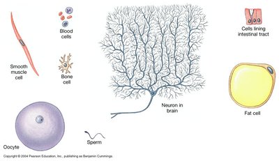
Cells are the fundamental units of life, performing all vital physiological functions. In the human body, cells are primarily eukaryotic, with specialized structures and functions. Prokaryotic cells (such as bacteria) and viruses are not considered part of the human cellular makeup but are important in pathology.
Eukaryotic cells: Have a nucleus and organelles; size ranges from 10–100 µm.
Prokaryotic cells: Lack a nucleus; smaller (1–10 µm); include bacteria.
Parasites: Eukaryotic, can be single-celled (protists) or multicellular (fungi, worms).
Viruses: Not cells; require host cells to replicate.
Cell Structure and Homeostasis
Each cell maintains homeostasis at the cellular level, and the coordinated action of many cells supports homeostasis at the tissue and organ levels. Cells are surrounded by extracellular (interstitial) fluid and are bounded by the plasma membrane.
Plasma membrane: Separates intracellular and extracellular environments, maintaining cellular homeostasis.
Cytoplasm: Includes cytosol (fluid) and organelles.
Nucleus: Contains genetic material (DNA).
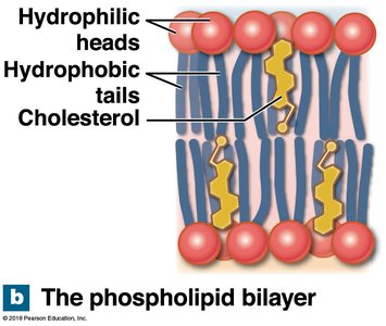
Plasma Membrane Structure
The plasma membrane is a dynamic, selectively permeable barrier composed of a phospholipid bilayer with embedded proteins, cholesterol, and carbohydrates.
Phospholipid bilayer: Hydrophilic phosphate heads face outward; hydrophobic fatty acid tails face inward.
Cholesterol: Stabilizes membrane fluidity.
Glycocalyx: Carbohydrate-rich area on the cell surface for recognition and protection.
Functions of the Plasma Membrane
Physical isolation: Maintains distinct internal and external environments.
Regulation of exchange: Controls entry/exit of ions, nutrients, and wastes.
Sensitivity and recognition: Responds to chemical signals; identifies cells as "self".
Structural support: Anchors cells and tissues.
Membrane Proteins and Carbohydrates
Transport proteins: Channels and carriers for movement of substances.
Receptors: Bind signaling molecules and trigger cellular responses.
Anchoring proteins: Attach the cell to other cells or the extracellular matrix.
Recognition proteins: Identify the cell to the immune system.
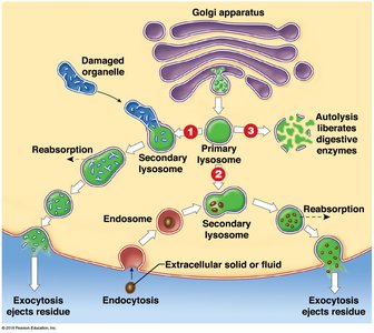
Membrane Flow and Organelle Dynamics
The cell membrane is part of a dynamic system involving the endoplasmic reticulum (ER), Golgi apparatus, and vesicles. Membrane components are synthesized, recycled, and transported via vesicular trafficking (endocytosis and exocytosis).
Rough ER (RER): Studded with ribosomes; synthesizes proteins.
Smooth ER (SER): Synthesizes lipids and carbohydrates.
Golgi apparatus: Modifies, sorts, and packages proteins and lipids.
Digestive Organelles
Proteasomes: Degrade damaged or abnormal proteins.
Peroxisomes: Break down lipids and toxins using enzymes.
Lysosomes: Vesicles containing digestive enzymes; responsible for autolysis (self-destruction of injured cells) and recycling cellular components.
Mitochondria
Mitochondria are the sites of aerobic respiration, producing ATP by metabolizing small organic molecules in the presence of oxygen. They are abundant in metabolically active cells, such as cardiac muscle cells.
Cellular respiration:
Nucleus and Genetic Control
Chromosomes: Store genetic information as DNA.
Gene expression: DNA is transcribed to mRNA, which is translated by ribosomes to synthesize proteins.
Proteins: Directly coded by DNA; enzymes produced by proteins synthesize lipids and carbohydrates.
Cell Cycle and Differentiation
All somatic cells derive from a fertilized egg and contain the same genes. Differentiation results from selective gene activation, producing specialized cells with limited capabilities. Adult stem cells retain the ability to divide and differentiate for tissue growth and repair.
Undifferentiated cells: Can become any cell type (pluripotent).
Differentiated cells: Specialized for specific functions.
The Tissue Level of Organization
Overview of Tissues
Tissues are collections of specialized cells and cell products that perform specific functions. Most organs contain all four primary tissue types:
Epithelial tissue
Connective tissue
Muscle tissue
Neural tissue
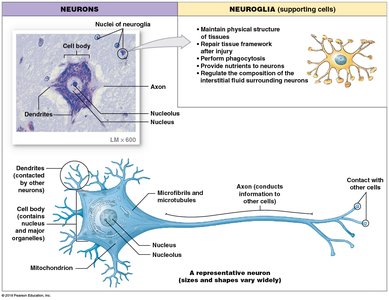
Neural Tissue
Neural tissue consists of neurons and supporting cells (neuroglia). It conveys information via electrical signals, integrates and interprets stimuli, and controls effectors such as muscles.
Neurons: Transmit electrical impulses.
Neuroglia: Support, protect, and nourish neurons.
Muscle Tissue
Muscle tissue is specialized for contraction and includes three types:
Skeletal muscle
Cardiac muscle
Smooth muscle
Connective Tissue
Connective tissue consists of specialized cells embedded in an extracellular matrix of fibers and fluid. It provides structural support, transports materials, protects organs, stores energy, and defends against pathogens.
Connective tissue proper: Includes fibroblasts, collagen, reticular, and elastic fibers (e.g., adipose, areolar, tendons, ligaments).
Fluid connective tissue: Blood and lymph.
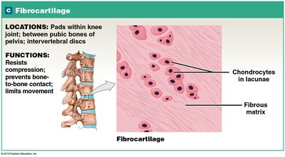
Supporting connective tissue: Cartilage and bone.
Epithelial Tissue
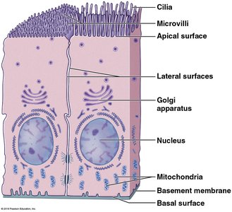
Epithelial tissue covers external surfaces, lines internal surfaces, and forms glands. It exhibits polarity, with an apical (free) surface and a basal surface attached to underlying connective tissue via the basal lamina.
Functions: Protection, absorption, secretion, sensation.
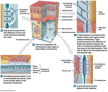
Specializations: Cilia (move materials), microvilli (increase surface area), tight junctions, gap junctions, desmosomes (cell connections).
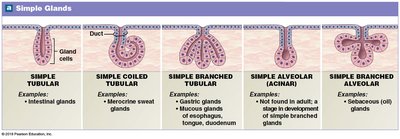
Glandular Epithelium
Glandular epithelia form exocrine and endocrine glands:
Exocrine glands: Secrete products through ducts onto epithelial surfaces (e.g., sweat, saliva).
Endocrine glands: Release hormones into interstitial fluid and blood.
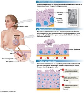
Modes of Glandular Secretion
Merocrine secretion: Product released by exocytosis (e.g., salivary glands).
Apocrine secretion: Involves loss of both product and cytoplasm (e.g., mammary glands).
Holocrine secretion: Destroys the cell as the product is released (e.g., sebaceous glands).
Summary Table: Four Basic Tissue Types
Tissue Type | Main Function | Examples |
|---|---|---|
Epithelial | Covering, lining, secretion, absorption | Skin, lining of GI tract, glands |
Connective | Support, binding, transport, energy storage | Bone, blood, fat, tendons |
Muscle | Contraction, movement | Skeletal muscle, heart, smooth muscle in organs |
Neural | Communication, control | Brain, spinal cord, nerves |
Additional info: This guide expands on the provided notes with definitions, examples, and academic context to ensure a comprehensive understanding of the cellular and tissue levels of organization in human anatomy and physiology.
