 Back
BackThe Cellular Level of Organization: Structure and Function of Eukaryotic Cells
Study Guide - Smart Notes
 Tailored notes based on your materials, expanded with key definitions, examples, and context.
Chapter 3: The Cellular Level of Organization
Introduction to Cells
Cells are the fundamental units of life in the human body. They are responsible for carrying out essential physiological functions and maintaining homeostasis at the cellular level. The collective activity of cells underpins the function of tissues, organs, and organ systems.
Definition: Cells are the smallest living units in the human body (eukaryotic cells).
Origin: All cells arise from the division of preexisting cells.
Function: Each cell maintains its own homeostasis, contributing to the overall stability of the organism.
Scale: The human body contains trillions of cells, outnumbered by microbial cells.
Plasma Membrane
Structure and Function
The plasma membrane, or cell membrane, forms the outer boundary of the cell and regulates the movement of substances into and out of the cell. It separates the intracellular fluid (ICF) from the extracellular fluid (ECF), maintaining distinct environments.
Composition: Primarily composed of a phospholipid bilayer and cholesterol.
Functions:
Physical separation of ICF and ECF.
Regulation of substance entry and exit.
Sensitivity: Contains receptors for environmental signals.
Support: Anchors cells to each other and to extracellular materials.
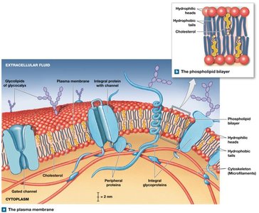
Phospholipid Bilayer
Hydrophilic heads: Face outward toward water in ECF and ICF.
Hydrophobic tails: Face inward, repelling water and ions.
Cholesterol: Stiffens the membrane, reducing fluidity and permeability.
Membrane Proteins
Integral proteins: Embedded within the membrane; some span the entire membrane (transmembrane proteins).
Peripheral proteins: Bound to the inner or outer surface of the membrane.
Eukaryotic Cell Organelles
Cytoplasm and Its Components
The cytoplasm includes all materials between the plasma membrane and the nuclear envelope. It consists of cytosol, organelles, and inclusions.
Cytosol (ICF): Jelly-like fluid containing water, nutrients, ions, proteins, and waste products.
Organelles: Specialized structures with specific functions.
Inclusions: Insoluble materials such as glycogen granules, lipid droplets, and pigment granules.
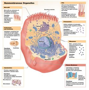
Nonmembranous Organelles
Nonmembranous organelles are not enclosed by membranes and are in direct contact with the cytosol. Examples include the cytoskeleton, centrioles, and ribosomes.
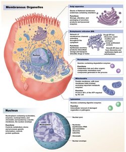
Membranous Organelles
Membranous organelles are surrounded by one or more phospholipid membranes, isolating them from the cytosol. These include the endoplasmic reticulum (ER), Golgi apparatus, lysosomes, peroxisomes, nucleus, and mitochondria.
Cytoskeleton
The cytoskeleton is an internal framework of proteins that provides structural support, strength, and flexibility to the cell. It consists of three types of fibers:
Microfilaments: Smallest fibers, composed of actin; provide mechanical strength and interact with myosin for muscle contraction.
Intermediate filaments: Provide strength and help maintain cell shape; stabilize organelle positions.
Microtubules: Largest fibers, made of tubulin; radiate from the centrosome, anchor organelles, and form the spindle apparatus during cell division.

Centrosome and Centrioles
Centrosome: Microtubule-organizing center near the nucleus.
Centrioles: Cylindrical structures within the centrosome; form the spindle apparatus during cell division.
Ribosomes
Function: Synthesize proteins.
Structure: Composed of small and large subunits containing rRNA and proteins.
Types: Free ribosomes (in cytosol) and fixed ribosomes (attached to RER).
Endoplasmic Reticulum (ER)
Structure: Network of membranes continuous with the nuclear envelope.
Functions: Synthesis of proteins, carbohydrates, and lipids; storage; transport; detoxification.
Types:
Smooth ER (SER): Lacks ribosomes; synthesizes lipids, steroid hormones, and stores Ca2+.
Rough ER (RER): Studded with ribosomes; synthesizes and folds proteins for export or membrane insertion.
Golgi Apparatus
Structure: Stack of flattened membranous discs (cisternae).
Functions: Modifies, sorts, and packages proteins and lipids for secretion or delivery to other organelles; forms lysosomes.
Lysosomes
Function: Contain digestive enzymes for breaking down macromolecules, recycling organelles, and destroying pathogens.
Autolysis: Self-destruction of damaged cells via lysosomal enzyme release.
Mitochondria
Function: Produce ATP through aerobic respiration.
Distribution: Number varies by cell type, reflecting energy demands (e.g., abundant in muscle cells, absent in RBCs).
Nucleus
Function: Control center of the cell; stores genetic information (DNA) and regulates protein synthesis.
Structure: Surrounded by a double membrane (nuclear envelope) with nuclear pores for molecular exchange.
Nucleolus: Site of rRNA synthesis and ribosome assembly.
Chromatin and Chromosomes: DNA is organized as chromatin in non-dividing cells and as chromosomes during cell division.
Protein Synthesis
Genetic Code and Central Dogma
The genetic code is the sequence of DNA nucleotides (A, T, C, G) that determines the amino acid sequence of proteins. The central dogma of molecular biology describes the flow of genetic information: DNA → mRNA → Protein.
Gene: A DNA sequence coding for a single protein.
Transcription: Synthesis of mRNA from a DNA template in the nucleus.
Translation: Synthesis of a polypeptide from an mRNA template at the ribosome.
Transcription
DNA is transcribed into mRNA, which can exit the nucleus.
mRNA is processed (splicing): introns are removed, exons are joined.
Alternative splicing allows one gene to code for multiple proteins.
Translation
mRNA binds to a ribosome.
tRNA molecules deliver amino acids, matching mRNA codons with complementary anticodons.
The amino acid sequence forms a polypeptide chain.
Transport Across the Plasma Membrane
Selective Permeability
The plasma membrane is selectively permeable, allowing some substances to pass freely while restricting others based on size, charge, shape, and solubility.
Passive and Active Transport
Passive transport: No energy required (e.g., diffusion, osmosis, facilitated diffusion).
Active transport: Requires energy (usually ATP) to move substances against their concentration gradients.
Diffusion
Movement of substances from high to low concentration (down the concentration gradient).
Simple diffusion: Lipid-soluble molecules and small gases (O2, CO2, H2O) cross the membrane directly.
Channel-mediated diffusion: Small water-soluble molecules and ions pass through protein channels.
Osmosis
Net diffusion of water across a selectively permeable membrane in response to solute concentration differences.
Water moves toward the solution with higher solute concentration (lower water concentration) until equilibrium is reached.
Tonicity
Isotonic: Equal solute concentrations; no net water movement.
Hypotonic: Lower solute concentration outside the cell; water enters, cell swells (possible lysis).
Hypertonic: Higher solute concentration outside the cell; water exits, cell shrivels (crenation).
Carrier-Mediated Transport
Involves specialized membrane proteins that transport specific substances.
Can be passive (facilitated diffusion) or active (requires ATP).
Symporters: Move two substances in the same direction.
Antiporters: Move two substances in opposite directions.
Sodium-potassium pump: Exchanges 3 Na+ out for 2 K+ in per ATP hydrolyzed; maintains cellular homeostasis.
Cell Life Cycle
Overview
Cell division is essential for growth, repair, and maintenance. It involves the production of two identical daughter cells from a single parent cell.
Mitosis: Division of somatic (body) cells.
Meiosis: Division producing gametes (sex cells).
Apoptosis: Programmed cell death.
Interphase
Period between cell divisions; includes G1 (growth), S (DNA replication), and G2 (protein synthesis) phases.
G0 phase: Non-dividing state (e.g., neurons, muscle cells).
Mitosis and Cytokinesis
Prophase: Chromosomes condense, nuclear envelope disintegrates, spindle fibers form.
Metaphase: Chromosomes align at the metaphase plate.
Anaphase: Sister chromatids separate and move to opposite poles.
Telophase: Nuclear membranes reform, chromosomes decondense.
Cytokinesis: Division of cytoplasm, forming two daughter cells.
Summary Table: Types of Membrane Transport
Type | Energy Required? | Direction | Example |
|---|---|---|---|
Simple Diffusion | No | High to Low | O2, CO2 |
Facilitated Diffusion | No | High to Low | Glucose transport |
Osmosis | No | Water: Low to High solute | Water movement |
Active Transport | Yes (ATP) | Low to High | Sodium-potassium pump |
Additional info: This guide expands on the provided slides with definitions, examples, and a summary table for clarity and exam preparation.
