 Back
BackThe Cellular Level of Organization: Structure and Function of the Plasma Membrane
Study Guide - Smart Notes
 Tailored notes based on your materials, expanded with key definitions, examples, and context.
Cellular Level of Organization
Introduction to Cells and Body Fluid Compartments
The cell is the fundamental unit of life, forming the basis for all physiological processes in the human body. Understanding the structure and function of cells, as well as the compartments in which body fluids are distributed, is essential for comprehending cellular interactions and homeostasis.
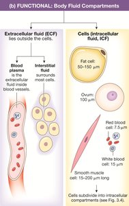
Intracellular fluid (ICF): Fluid within cells, making up about 66% of total body water.
Extracellular fluid (ECF): Fluid outside cells, including interstitial fluid (25%) and plasma (8%).
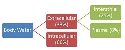
Key Point: The plasma membrane separates the ICF from the ECF, maintaining distinct environments essential for cellular function.
The Plasma Membrane
Structure and Composition
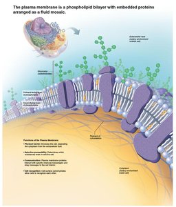
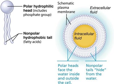
The plasma membrane, also known as the cell membrane, is a dynamic structure that serves as a selective barrier between the cell's internal and external environments. It is described by the fluid mosaic model, reflecting its flexible, ever-changing composition of lipids, proteins, and carbohydrates.
Phospholipid bilayer: Forms the basic structure, with hydrophilic (water-loving) heads facing outward and hydrophobic (water-fearing) tails facing inward.
Cholesterol: Stabilizes membrane fluidity and integrity.
Glycolipids and glycoproteins: Contribute to the glycocalyx, a "sugar coating" involved in cell recognition.
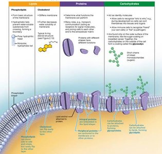
Membrane Proteins
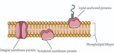
Membrane proteins are critical for communication, transport, and structural support. They are classified as:
Integral proteins: Span the membrane; function as channels, carriers, receptors, or enzymes.
Peripheral proteins: Loosely attached to the membrane; involved in support, signaling, and cell shape.
Functions of Membrane Proteins
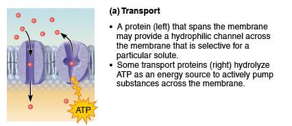
Transport: Move substances across the membrane (channels, pumps).
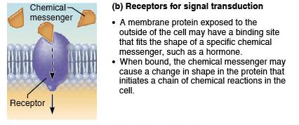
Receptors for signal transduction: Bind chemical messengers and initiate cellular responses.
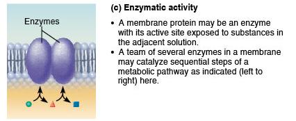
Enzymatic activity: Catalyze metabolic reactions.
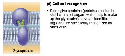
Cell-cell recognition: Glycoproteins serve as identification tags.
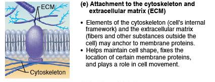
Attachment to cytoskeleton and ECM: Maintain cell shape and stabilize membrane proteins.
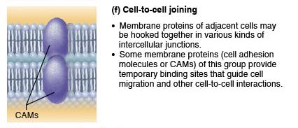
Cell-to-cell joining: Form intercellular junctions for tissue integrity.
Cell Junctions
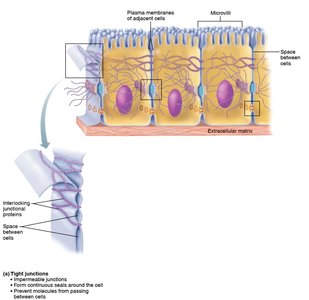
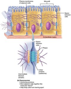
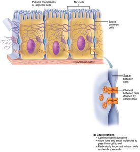
Cell junctions are specialized structures that connect adjacent cells, providing communication and structural integrity. The three main types are:
Tight junctions: Create impermeable seals to prevent passage of molecules between cells.
Desmosomes: Anchor cells together, providing mechanical strength.
Gap junctions: Allow direct communication between cells via connexons (protein channels).
Membrane Transport Mechanisms
Passive Transport
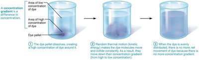
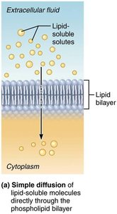
Passive transport does not require cellular energy (ATP) and relies on the movement of substances down their concentration gradients.
Simple diffusion: Nonpolar, lipid-soluble molecules move directly through the bilayer.
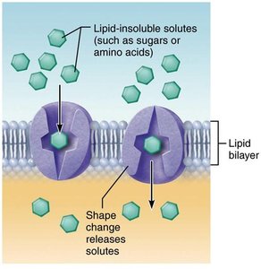
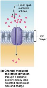
Facilitated diffusion: Polar or charged molecules move via protein carriers or channels.
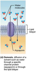
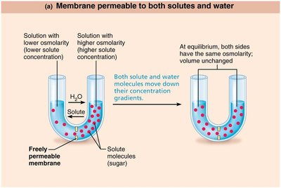
Osmosis: Diffusion of water across a selectively permeable membrane.
Osmolarity and Tonicity
Osmolarity is the measure of solute concentration. Tonicity describes how a solution affects cell volume:
Isotonic: No net water movement; cell volume remains unchanged.
Hypertonic: Water leaves the cell; cell shrinks (crenation).
Hypotonic: Water enters the cell; cell swells and may lyse.
Active Transport
Active transport requires energy (ATP) to move substances against their concentration gradients. It is essential for maintaining cellular homeostasis.
Primary active transport: Direct use of ATP to transport molecules (e.g., Na+/K+ ATPase pump).
Secondary active transport: Indirect use of ATP; uses gradients established by primary active transport to move other substances (symporters and antiporters).
Membrane Potential
Resting Membrane Potential (RMP)
The resting membrane potential is the voltage difference across the plasma membrane in resting cells, typically ranging from –50 to –100 mV. It is primarily established by the differential distribution of K+ and Na+ ions, maintained by the Na+/K+ ATPase pump.
K+ diffusion: Outward movement creates a negative charge inside the cell.
Na+ diffusion: Inward movement slightly offsets the negative charge.
Na+/K+ pump: Maintains gradients by pumping 3 Na+ out and 2 K+ in per ATP hydrolyzed.
Clinical and Applied Context
Understanding the plasma membrane and its transport mechanisms is crucial for interpreting laboratory values, managing fluid and electrolyte balance, and understanding drug actions and cellular responses in health sciences.
Example: Placing a red blood cell in a hypotonic solution (e.g., 0.1% NaCl) causes water to enter the cell, leading to swelling and possible lysis. In a hypertonic solution (e.g., 5% NaCl), water leaves the cell, causing it to shrink.
Additional info: This knowledge is foundational for fields such as nursing, pharmacy, and medical technology, where cellular physiology underpins clinical practice and patient care.
