 Back
The Central and Peripheral Nervous System: Structure and Function
Study Guide - Smart Notes
 Tailored notes based on your materials, expanded with key definitions, examples, and context.
The Nervous System Overview
Central Nervous System (CNS) and Peripheral Nervous System (PNS)
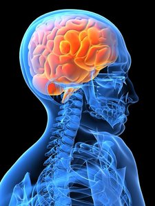
The nervous system is divided into the Central Nervous System (CNS)—comprising the brain and spinal cord—and the Peripheral Nervous System (PNS), which includes all neural tissue outside the CNS. The CNS integrates sensory information and coordinates body responses, while the PNS connects the CNS to limbs and organs.
Neural Tissue Structure
Multipolar Neurons
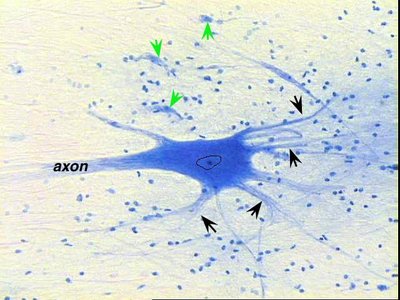
Multipolar neurons are the most common type of neuron in the CNS. They have multiple dendrites and a single axon, allowing them to integrate a large amount of information from other neurons.
Cell body (soma): Contains the nucleus and organelles.
Dendrites: Receive incoming signals.
Axon: Transmits electrical impulses away from the cell body.
Spinal Cord Structure and Function
Gross Anatomy of the Spinal Cord
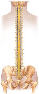
The spinal cord is a cylindrical structure that extends from the foramen magnum to the level of the first or second lumbar vertebra. It is protected by vertebrae, meninges, and cerebrospinal fluid (CSF).
Cervical and lumbar enlargements: Regions where nerves serving the limbs arise.
Conus medullaris: The cone-shaped inferior end of the spinal cord.
Cauda equina: A bundle of nerve roots at the lower end of the vertebral canal.
Filum terminale: A fibrous extension anchoring the spinal cord to the coccyx.
Cross-Sectional Anatomy of the Spinal Cord
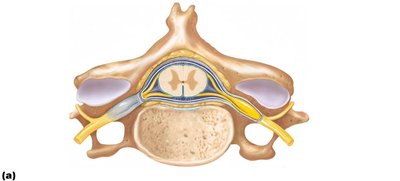
In cross-section, the spinal cord shows a central region of gray matter (containing neuron cell bodies) surrounded by white matter (containing myelinated axons).
Dorsal horn: Contains interneurons receiving sensory input.
Ventral horn: Contains somatic motor neuron cell bodies.
Lateral horn: (only in thoracic and lumbar regions) Contains autonomic motor neuron cell bodies.
Dorsal root: Contains sensory neuron axons; dorsal root ganglion contains their cell bodies.
Ventral root: Contains motor neuron axons.
Spinal nerve: Formed by the fusion of dorsal and ventral roots; a mixed nerve carrying both sensory and motor fibers.
Spinal Nerves and Plexuses
Spinal Nerves
There are 31 pairs of spinal nerves, all of which are mixed nerves (containing both sensory and motor fibers). They are named according to their point of origin from the spinal cord:
8 pairs of cervical nerves (C1–C8)
12 pairs of thoracic nerves (T1–T12)
5 pairs of lumbar nerves (L1–L5)
5 pairs of sacral nerves (S1–S5)
1 pair of coccygeal nerves (C0)
After exiting the vertebral column, each spinal nerve divides into three branches:
Dorsal ramus: Supplies the posterior body trunk.
Ventral ramus: Supplies the rest of the trunk and limbs.
Meningeal branch: Reenters the vertebral canal to innervate meninges and blood vessels.
Nerve Plexuses
Except for the thoracic region (T2–T12), ventral rami of spinal nerves form nerve plexuses—interlacing networks of nerves. These plexuses ensure that each limb muscle is innervated by more than one spinal nerve, providing redundancy in case of injury.
Cervical plexus (C1–C4): Innervates skin of neck, shoulder, and diaphragm (via the phrenic nerve).
Brachial plexus (C5–C8, T1): Innervates the upper limb.
Lumbosacral plexus (L1–S4): Innervates the leg, buttocks, and abdomen. Major nerves include the femoral, obturator, and sciatic nerves.
Protective Structures of the CNS
Meninges
The meninges are three connective tissue membranes that cover and protect the CNS, enclose venous sinuses, contain cerebrospinal fluid (CSF), and form partitions within the skull.
Dura mater: Outermost, tough layer.
Arachnoid mater: Middle, web-like layer.
Pia mater: Innermost, delicate layer adhering to the CNS surface.
Spaces associated with the meninges include the epidural space (contains fat), subdural space, and subarachnoid space (contains CSF).
Functional Organization of the Spinal Cord
Gray and White Matter Organization
The spinal cord's gray matter is organized into horns (dorsal, ventral, and lateral), each with specific functional roles:
Dorsal horn: Receives somatic and visceral sensory input.
Ventral horn: Contains somatic motor neurons.
Lateral horn: Contains autonomic (visceral motor) neurons (only in thoracic and lumbar regions).
The white matter is organized into funiculi (dorsal, lateral, ventral) containing ascending and descending nerve tracts.
Summary Table: Spinal Cord and Spinal Nerves
Structure | Function | Location/Notes |
|---|---|---|
Cervical enlargement | Nerves to upper limbs | Neck region |
Lumbar enlargement | Nerves to lower limbs | Lower back |
Conus medullaris | Inferior end of spinal cord | L1–L2 vertebral level |
Cauda equina | Nerve roots for lower limbs | Inferior to conus medullaris |
Filum terminale | Anchors spinal cord | Extends to coccyx |
Dorsal root | Sensory input to cord | Contains sensory axons |
Ventral root | Motor output from cord | Contains motor axons |
Spinal nerve | Mixed (sensory & motor) | Fusion of dorsal & ventral roots |
