 Back
BackThe Central Nervous System: Structure and Function of the Brain
Study Guide - Smart Notes
 Tailored notes based on your materials, expanded with key definitions, examples, and context.
The Midbrain (Mesencephalon)
Cross Section and Major Structures
The midbrain is a portion of the brainstem that connects the hindbrain to the forebrain. It contains several important structures involved in motor control, sensory processing, and consciousness.
Tegmentum: Connects to the cerebellum and helps control fine movements through the red nucleus, which is rich in blood vessels.
Substantia nigra: Sends inhibitory signals to the basal ganglia and thalamus; degeneration leads to tremors and Parkinson's disease.
Cerebral crus: Connects the cerebrum to the pons.
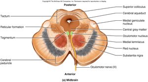
Superior and Inferior Colliculi (Tectum)
The tectum of the midbrain contains four nuclei known as the corpora quadrigemina:
Superior colliculus: Involved in tracking moving objects, blinking, and pupillary and head-turning reflexes.
Inferior colliculus: Responsible for reflex turning of the head in response to sound.
Reticular Formation
The reticular formation is a network of gray matter scattered throughout the pons, midbrain, and medulla. It plays a crucial role in regulating balance, posture, sleep, and consciousness.
Relays information from eyes and ears to the cerebellum.
Contains gaze centers and central pattern generators.
Regulates sleep and conscious attention; injury can lead to irreversible coma.
Includes cardiac and vasomotor centers, and is the origin of descending analgesic pathways for pain modulation.
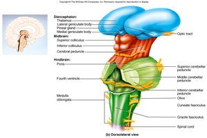
The Forebrain
Cerebrum and Diencephalon
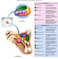
The forebrain consists of the cerebrum and the diencephalon. The diencephalon includes the thalamus, hypothalamus, and epithalamus.
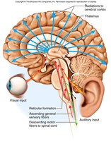
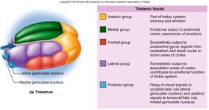
Thalamus: Makes up 4/5 of the diencephalon and contains over 23 nuclei. It is the main relay station for sensory information to the cerebral cortex and is involved in emotional and memory functions (limbic system).
Hypothalamus: Forms the walls and floor of the third ventricle. It regulates hormone secretion, autonomic nervous system control, thermoregulation, hunger, thirst, sleep, circadian rhythms, memory, and emotional behavior.
Epithalamus (Pineal Gland): Contains the pineal gland, which secretes serotonin and melatonin, and the habenula, which connects the limbic system to the midbrain.
Cerebrum: Gross Anatomy and Lobes
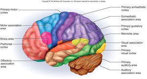
Structure and Functional Areas
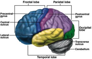
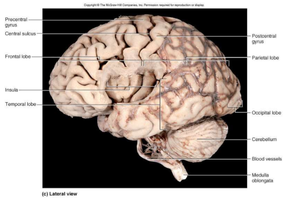
The cerebrum is the largest part of the brain, with a highly folded cerebral cortex (about 3 mm thick) to increase surface area. It is divided into several lobes, each with specialized functions:
Frontal lobe: Voluntary motor functions, planning, mood, smell, and social judgment.
Parietal lobe: Receives and integrates sensory information.
Occipital lobe: Visual processing center.
Temporal lobe: Hearing, smell, learning, memory, and emotional behavior.
Insula: Understanding language, taste, and visceral sensory information.
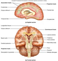
White Matter Tracts of the Cerebrum
Types of Tracts
Most of the cerebrum consists of white matter, which contains myelinated nerve fibers organized into three types of tracts:
Projection tracts: Connect the cerebrum to lower brain and spinal cord centers.
Commissural tracts: Cross between hemispheres (e.g., corpus callosum, anterior and posterior commissures).
Association tracts: Connect different regions within the same hemisphere.
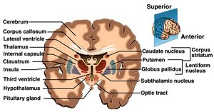
Basal Nuclei and Limbic System
Basal Nuclei
The basal nuclei are masses of gray matter deep within the cerebral hemispheres. They receive input from the substantia nigra and motor cortex and are involved in motor control and inhibition of tremors.
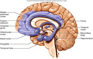
Limbic System
The limbic system is a loop of cortical structures surrounding the deep brain, including the amygdala, hippocampus, fornix, and cingulate gyrus. It is essential for emotion and memory processing.
Amygdala: Important for emotional responses.
Hippocampus: Critical for memory formation.
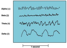
Electroencephalogram (EEG) and Brain Waves
Types of Brain Waves
An electroencephalogram (EEG) records voltage changes from postsynaptic potentials in the cerebral cortex. There are four main types of brain waves, each associated with different states of consciousness:
Alpha waves: Awake and resting with eyes closed.
Beta waves: Eyes open, performing mental tasks.
Theta waves: Occur during sleep or emotional stress.
Delta waves: Deep sleep.
Sleep and Sleep Stages
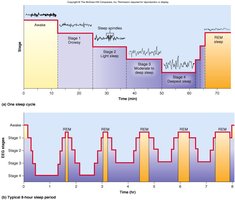
Non-REM and REM Sleep
Sleep is a temporary state of unconsciousness with distinct stages:
Non-REM sleep: Four stages, progressing from light to deep sleep, with restorative effects.
REM sleep: Characterized by rapid eye movements, increased vital signs, vivid dreams, and EEG patterns similar to wakefulness. REM periods become longer and more frequent as the night progresses.
Cognition and Brain Lesions
Mental Processes and Effects of Lesions
Cognition includes awareness, perception, thinking, knowledge, and memory. The association areas of the brain integrate sensory and motor information. Lesions in different lobes can cause specific deficits:
Parietal lobe: Contralateral neglect syndrome (unawareness of one side of the body).
Temporal lobe: Agnosia (inability to recognize objects), prosopagnosia (inability to recognize faces).
Frontal lobe: Personality changes, inability to plan or execute appropriate behavior.
Memory and Emotion
Memory Formation and Emotional Processing
Memory involves learning, storing, and forgetting information. The hippocampus is essential for forming new memories, while the cerebellum helps with motor skills and the amygdala with emotional memory. The prefrontal cortex controls emotional expression, while emotions are generated in the hypothalamus and amygdala.
Somesthetic Sensation and Sensory Homunculus
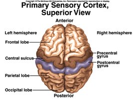
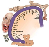
Somatosensory Cortex
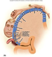
Somesthetic sensations (touch, pain, pressure, etc.) are processed in the postcentral gyrus of the parietal lobe. The sensory homunculus illustrates that the amount of cortex dedicated to each body part is proportional to its sensitivity.
Functional Regions of the Cerebral Cortex
Special Senses and Association Areas
Special senses (smell, taste, vision, hearing, equilibrium) are processed in specific regions of the cortex. Association areas interpret sensory information and are responsible for higher-order processing such as recognition and memory.
Motor Control and Motor Homunculus
Motor Cortex Organization
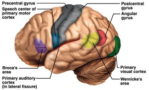
The intention to move begins in the premotor area of the frontal lobe. The precentral gyrus (primary motor area) sends signals to the spinal cord, controlling muscles on the opposite side of the body. The motor homunculus shows the proportion of cortex devoted to different muscle groups, reflecting the degree of fine control required.

Language Centers
Broca's and Wernicke's Areas
Language involves several brain regions:
Wernicke's area: Recognition of spoken and written language, creation of speech plans.
Broca's area: Generates motor programs for speech production.
Affective language area: Adds emotional content to speech; lesions cause flat, emotionless speech (aprosodia).
Aphasia
Aphasia is any language deficit due to lesions in the language-dominant hemisphere:
Broca's aphasia: Nonfluent, slow speech with limited vocabulary.
Wernicke's aphasia: Fluent but nonsensical speech.
Anomic aphasia: Normal speech and understanding, but inability to make sense of text or pictures.
Cerebral Lateralization
Hemispheric Specialization
The two cerebral hemispheres are specialized for different functions:
Left hemisphere (categorical): Language, sequential and analytical reasoning, math, and science.
Right hemisphere (representational): Holistic perception, spatial relationships, imagination, music, and artistic skill.
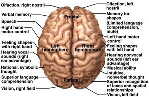
Lateralization is correlated with handedness and develops with age. Females tend to have more communication between hemispheres.
Disorders of the Central Nervous System
Common CNS Disorders
Cerebral palsy: Muscular incoordination due to brain damage during fetal development.
Concussion: Brain injury from a blow to the head, causing loss of consciousness and sensory disturbances.
Encephalitis: Brain inflammation due to infection, leading to neuronal degeneration and possible death.
Epilepsy: Sudden, massive neuronal discharge causing seizures.
Migraine headache: Severe, recurring headaches with associated symptoms.
Schizophrenia: Thought disorder involving delusions.
