 Back
BackThe Central Nervous System: Structure and Function
Study Guide - Smart Notes
 Tailored notes based on your materials, expanded with key definitions, examples, and context.
The Central Nervous System (CNS)
Overview and Major Functions
The central nervous system (CNS) consists of the brain and spinal cord. It is responsible for integrating sensory information, coordinating movement, maintaining homeostasis, and supporting higher mental functions such as language, learning, and memory.
Motor functions: Initiate muscle contraction and gland secretion (in conjunction with the peripheral nervous system, PNS).
Sensory functions: Interpret sensations from inside and outside the body (PNS input, CNS processing).
Integrative functions: Decision-making, planning, and monitoring movement, as well as maintaining homeostasis and higher mental activities.
Basic Structure of the Brain and Spinal Cord
Brain Divisions and Their Functions
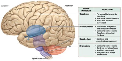
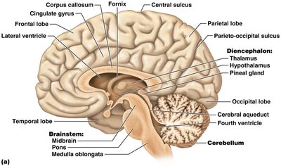
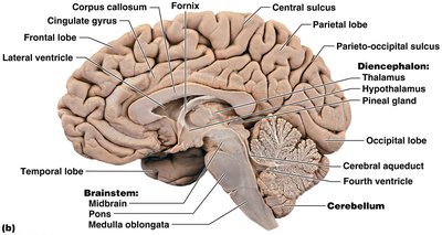
The brain is a soft, whitish-gray organ located in the cranial cavity and is continuous with the spinal cord. It is composed mostly of nervous tissue, with some epithelial and connective tissue. The brain is divided into four main regions, each with specialized functions:
Brain Division | Function |
|---|---|
Cerebrum | Performs higher mental functions, interprets sensory stimuli, plans and initiates movement |
Diencephalon | Processes, integrates, and relays information; maintains homeostasis; regulates biological rhythms |
Cerebellum | Monitors and coordinates movement |
Brainstem | Maintains homeostasis, controls certain reflexes, monitors movement, integrates and relays information |
White and Gray Matter
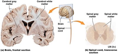
Both the brain and spinal cord contain white and gray matter:
White matter: Composed of myelinated axons; forms tracts that carry information.
Gray matter: Contains cell bodies, dendrites, and unmyelinated axons; forms nuclei and the cerebral cortex.
The Cerebrum
Surface Features: Gyri and Sulci
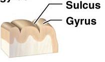
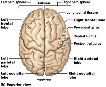
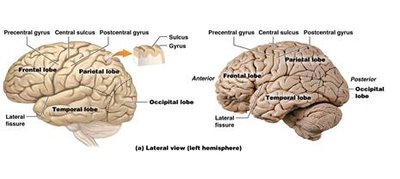
The surface of the cerebrum is highly folded, increasing its surface area. The folds are called gyri (ridges) and sulci (shallow grooves).
Lobes and Major Landmarks
Each cerebral hemisphere is divided into five lobes:
Frontal lobe: Motor functions, complex mental functions
Parietal lobe: Sensory processing
Temporal lobe: Hearing
Occipital lobe: Vision
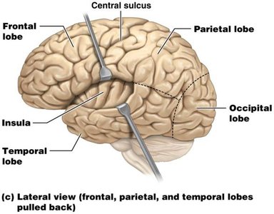
Insula: Taste and visceral sensation
Major fissures and gyri include the longitudinal fissure (separates hemispheres), central sulcus, precentral gyrus (primary motor cortex), and postcentral gyrus (primary somatosensory cortex).
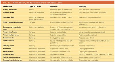
Cerebral Cortex: Functional Areas
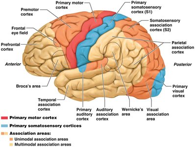
The cerebral cortex is the outer layer of gray matter covering the cerebral hemispheres. It is responsible for conscious processes such as planning movement, interpreting sensory information, and higher cognitive functions. The cortex is divided into three main functional areas:
Motor areas: Control voluntary movement (e.g., primary motor cortex in the precentral gyrus).
Sensory areas: Receive and process sensory input (e.g., primary somatosensory cortex in the postcentral gyrus, visual cortex in the occipital lobe).
Association areas: Integrate information for complex functions (e.g., prefrontal cortex for behavior and personality, Broca's and Wernicke's areas for language).
Area Name | Type of Cortex | Location | Function |
|---|---|---|---|
Primary motor cortex | Motor | Precentral gyrus of frontal lobe | Plans and executes movement |
Premotor cortex | Unimodal association | Widespread throughout lateral and medial frontal lobe | Plans and executes complex movement |
Frontal eye fields | Unimodal association | Anterior to the premotor cortex | Back-and-forth eye movements |
Primary somatosensory cortex | Sensory | Postcentral gyrus of parietal lobe | Interprets incoming somatic sensory information |
Somatosensory association cortex | Unimodal association | Posterior to the primary somatosensory cortex in the parietal lobe | Integrates somatic sensory information |
Primary visual cortex | Sensory | Occipital lobe | Interprets and processes visual stimuli |
Visual association area | Unimodal association | Occipital lobe | Processes visual stimuli |
Primary auditory cortex | Sensory | Superior temporal lobe | Processes auditory stimuli |
Auditory association cortex | Unimodal association | Superior temporal lobe | Processes auditory stimuli |
Vestibular areas | Sensory | Parietal and temporal lobes | Processes stimuli relating to equilibrium and balance |
Olfactory cortex | Sensory | Temporal lobe | Processes smell stimuli |
Broca's area | Multimodal association | Superolateral frontal lobe | Language production |
Wernicke's area | Multimodal association | Superolateral temporal lobe | Language comprehension |
Prefrontal cortex | Multimodal association | Anterior frontal lobe | Planning, personality, higher cognitive functions |
Parietal association cortex | Multimodal association | Widespread in the cerebral lobe | Spatial awareness and attention |
Temporal association cortex | Multimodal association | Widespread in the temporal lobe | Recognition and association |
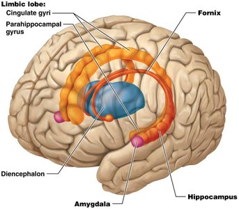
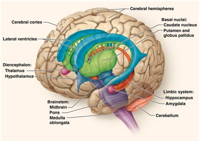
Basal Nuclei and Limbic System
Basal nuclei are masses of gray matter deep within each hemisphere, involved in movement regulation and inhibition of involuntary movements. The limbic system includes the limbic lobe, hippocampus, and amygdala, and is involved in memory, learning, emotion, and behavior.
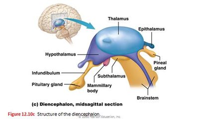
The Diencephalon
Major Components and Functions
The diencephalon is located centrally in the brain, between the hemispheres and above the brainstem. It consists of four main parts:
Thalamus: Gateway for sensory information to the cerebral cortex (except smell).
Hypothalamus: Regulates the autonomic nervous system, sleep/wake cycle, thirst, hunger, and body temperature; secretes hormones that regulate the pituitary gland.
Epithalamus: Contains the pineal gland, which secretes melatonin for sleep/wake regulation.
Subthalamus: Functionally connected with basal nuclei to control movement.
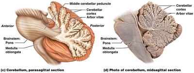
The Cerebellum
Structure and Function
The cerebellum is located inferior to the occipital lobe and is responsible for coordinating voluntary movement and maintaining balance. Its white matter forms the arbor vitae, a tree-like structure visible in section.
The Brainstem
Major Regions and Functions
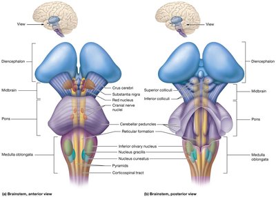
The brainstem is one of the oldest parts of the brain and is vital for survival. It includes the midbrain, pons, and medulla oblongata, and is responsible for basic involuntary processes such as breathing, heart rate, and reflexes.
Midbrain: Involved in visual and auditory reflexes; contains the substantia nigra, which produces dopamine and works with basal nuclei to control movement.
Pons: Regulates movement, breathing, reflexes, and sleep/arousal functions.
Medulla oblongata: Controls vital functions such as breathing and heart rate.
Protection of the Brain and Spinal Cord
Meninges, CSF, and Blood-Brain Barrier
The CNS is protected by three main features:
Cranial meninges: Three layers of membranes (dura mater, arachnoid mater, pia mater) surround the brain and spinal cord.
Cerebrospinal fluid (CSF): Bathes the brain and spinal cord, cushions them, and helps maintain temperature and remove wastes.
Blood-brain barrier: Prevents many substances from entering the brain from the blood.
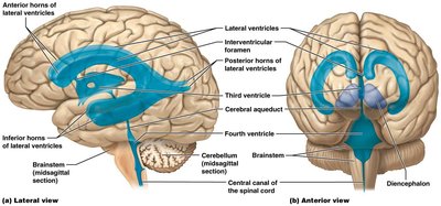
Ventricles and CSF Circulation
The brain contains four ventricles (lateral, third, and fourth) filled with CSF, which is produced by the choroid plexuses and reabsorbed by arachnoid villi. CSF circulates through the ventricles and central canal of the spinal cord.
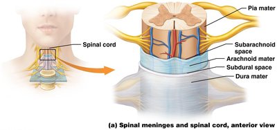
Spinal Cord Structure and Protection
Spinal Meninges and Spaces
The spinal cord is protected by meninges similar to those of the brain. Key spaces include:
Epidural space: Between dura mater and vertebral wall; contains veins and adipose tissue.
Subarachnoid space: Between arachnoid and pia mater; filled with CSF.

Summary Table: Major Brain Structures and Functions
Division | Main Function |
|---|---|
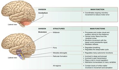
Cerebellum | Coordinates ongoing voluntary movement to reduce motor error |
Brainstem: Midbrain | Processes and routes visual and auditory stimuli to the thalamus; carries motor fibers from the cerebral cortex; monitors movement with the basal nuclei; mediates reflexes |
Brainstem: Pons | Regulates movement, breathing, reflexes, and sleep/wake cycle |
Brainstem: Medulla oblongata | Regulates many autonomic functions |
Brainstem: Reticular formation | Involved in sleep and arousal, pain transmission, and mood regulation |
All regions | Contain tracts of white matter involved in movement and sensation |
Additional info: The CNS is essential for integrating sensory input, coordinating voluntary and involuntary actions, and supporting cognition, emotion, and memory. Damage to any part of the CNS can result in significant functional deficits, highlighting the importance of its protection and maintenance.
