 Back
BackThe Central Nervous System: Structure and Function
Study Guide - Smart Notes
 Tailored notes based on your materials, expanded with key definitions, examples, and context.
The Central Nervous System (CNS)
Overview and Cephalization
The central nervous system (CNS) is composed of the brain and spinal cord. Cephalization refers to the evolutionary trend toward increasing concentration of nervous tissue and sensory organs at the anterior end of the body, culminating in the highly developed human brain.
Cephalization: Elaboration of the anterior CNS, with increased neuron numbers in the head region.
Brain Structure: The brain consists of wrinkled, pinkish-gray tissue, including the cerebral hemispheres, cerebellum, and brain stem.
Development and Organization of the CNS
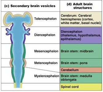
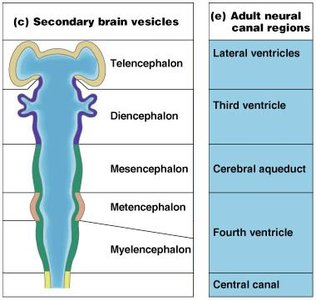
Neural Canal Regions and Brain Vesicles
The adult brain structures and neural canal regions are derived from embryonic brain vesicles. These vesicles give rise to specific adult brain regions and associated ventricles.
Telencephalon: Forms the cerebral hemispheres (cortex, white matter, basal nuclei) and lateral ventricles.
Diencephalon: Forms the thalamus, hypothalamus, epithalamus, and third ventricle.
Mesencephalon: Forms the midbrain and cerebral aqueduct.
Metencephalon: Forms the pons and cerebellum, associated with the fourth ventricle.
Myelencephalon: Forms the medulla oblongata and part of the fourth ventricle.
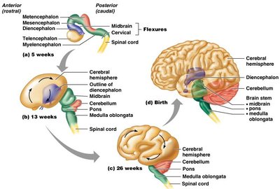
Brain Development and Flexures
During embryonic development, the brain undergoes flexures and expansion, resulting in the complex adult structure. The neural tube forms primary and secondary brain vesicles, which differentiate into the mature brain regions.
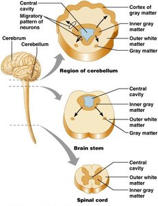
Basic Pattern of the CNS
The CNS exhibits a basic pattern of central gray matter surrounded by white matter. In the brain, additional gray matter forms the cortex and nuclei.
Spinal Cord: Central cavity with inner gray matter and outer white matter.
Brain Stem: Similar to spinal cord but with scattered gray matter nuclei.
Cerebellum and Cerebrum: Have additional outer gray matter (cortex).
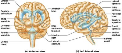
Ventricles of the Brain
Structure and Function
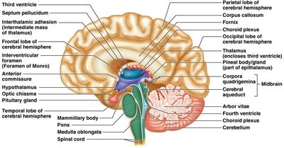
The brain contains interconnected cavities called ventricles, which are filled with cerebrospinal fluid (CSF). These include the paired lateral ventricles, third ventricle, and fourth ventricle.
Lateral Ventricles: Located in the cerebral hemispheres.
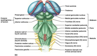
Third Ventricle: Located in the diencephalon.
Fourth Ventricle: Located dorsal to the pons and medulla.
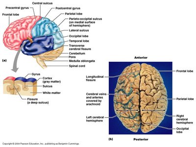
Cerebrum: Structure and Functional Areas
Lobes, Gyri, and Sulci
The cerebrum is divided into right and left hemispheres, each containing lobes separated by sulci and fissures. The surface features gyri (ridges) and sulci (shallow grooves).
Lobes: Frontal, parietal, temporal, occipital, and insula.
Major Sulci: Central sulcus, parieto-occipital sulcus, lateral sulcus.
Fissures: Longitudinal fissure separates hemispheres.
Cerebral Cortex
The cerebral cortex is the outer layer of gray matter, responsible for sensation, communication, memory, understanding, and voluntary movements. Each hemisphere controls the opposite side of the body (contralateral control).
Functional Areas: Motor areas (voluntary movement), sensory areas (conscious sensation), association areas (integration of information).
Lateralization: Each hemisphere has specialized functions (e.g., left for language, right for spatial skills).
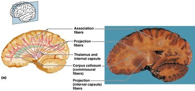
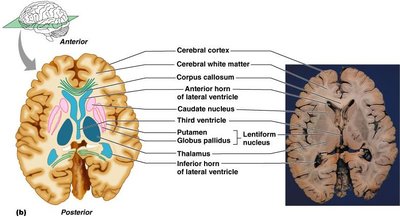
Cerebral White Matter and Basal Nuclei
White Matter Fiber Tracts
Cerebral white matter consists of myelinated fibers responsible for communication within the brain and with lower CNS centers.
Commissures: Connect corresponding gray areas of the two hemispheres (e.g., corpus callosum).
Association Fibers: Connect different parts of the same hemisphere.
Projection Fibers: Connect the cerebrum with lower brain or spinal cord centers.
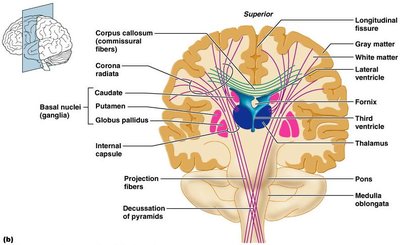
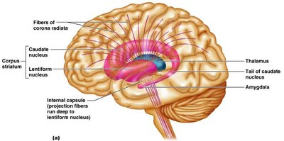
Basal Nuclei
Basal nuclei are clusters of gray matter deep within the cerebral white matter. They are involved in regulating movement and cognition.
Components: Caudate nucleus, putamen, globus pallidus (lentiform nucleus).
Functions: Influence muscular activity, regulate attention and cognition, inhibit unnecessary movements.
Diencephalon
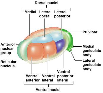
Thalamus
The thalamus is a paired, egg-shaped structure that forms the superolateral walls of the third ventricle. It acts as a relay station for sensory and motor signals to the cerebral cortex.
Nuclei: Anterior, ventral, dorsal, and posterior groups.
Function: Sorts, edits, and relays sensory information; involved in sensation, motor activities, learning, and memory.
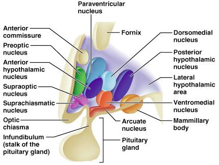
Hypothalamus
The hypothalamus lies below the thalamus and forms the inferolateral walls of the third ventricle. It is the main visceral control center, regulating homeostasis, endocrine function, and autonomic activities.
Key Structures: Mammillary bodies (olfactory relay), infundibulum (connects to pituitary gland).
Functions: Regulates body temperature, hunger, thirst, sleep, emotional responses, and hormone secretion.
Epithalamus
The epithalamus forms the roof of the third ventricle and includes the pineal gland, which secretes melatonin to regulate sleep-wake cycles and mood. The choroid plexus in this region produces cerebrospinal fluid.
Brain Stem
Regions and Functions
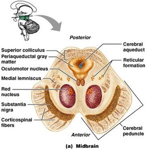
The brain stem consists of the midbrain, pons, and medulla oblongata. It controls automatic behaviors necessary for survival and serves as a conduit for tracts between higher and lower brain centers.
Midbrain: Contains cerebral peduncles, cerebral aqueduct, and nuclei for cranial nerves III and IV.
Pons: Connects higher brain centers with the spinal cord and relays impulses between the motor cortex and cerebellum.
Medulla Oblongata: Contains cardiovascular and respiratory centers, and is the site of decussation of pyramidal tracts.

The Cerebellum
Structure and Function
The cerebellum is located dorsal to the pons and medulla and is responsible for coordinating voluntary movements, balance, and posture. It also plays a role in cognitive functions such as language and problem-solving.
Hemispheres: Two, connected by the vermis.
Lobes: Anterior, posterior, and flocculonodular.
Arbor Vitae: Distinctive tree-like pattern of white matter.
Functional Brain Systems
Limbic System
The limbic system is involved in emotion, motivation, and memory. It includes structures such as the amygdala, hippocampus, and parts of the diencephalon.
Amygdala: Processes emotions like fear and anger.
Cingulate Gyrus: Involved in emotional expression and conflict resolution.
Hippocampus: Converts short-term memory to long-term memory.
Reticular Formation
The reticular formation is a network of neurons in the brain stem that regulates arousal, consciousness, and motor functions. The reticular activating system (RAS) keeps the cortex alert and filters sensory input.
Protection of the Brain
Meninges
The brain is protected by three connective tissue membranes: dura mater, arachnoid mater, and pia mater. These layers cover and protect the CNS, enclose blood vessels, and contain cerebrospinal fluid.
Dura Mater: Tough, outermost layer with periosteal and meningeal layers.
Arachnoid Mater: Middle layer with subarachnoid space filled with CSF.
Pia Mater: Delicate, innermost layer adhering to the brain surface.
Cerebrospinal Fluid (CSF)
CSF is a clear, watery fluid that cushions the brain, provides buoyancy, removes waste, and transports nutrients. It is produced by the choroid plexuses and circulates through the ventricles and subarachnoid space.
Formation: By filtration of blood plasma at the choroid plexuses.
Circulation: Flows through ventricles, subarachnoid space, and is absorbed into venous blood via arachnoid villi.
Blood-Brain Barrier
The blood-brain barrier is a selective barrier that protects the brain from harmful substances in the blood while allowing essential nutrients to pass. It is formed by endothelial cells, a thick basal lamina, and astrocyte end-feet.
The Spinal Cord
Structure and Function
The spinal cord extends from the foramen magnum to L1 or L2 and provides two-way communication between the brain and body. It is protected by bone, meninges, and CSF.
Conus Medullaris: Terminal end of the spinal cord.
Filum Terminale: Fibrous extension anchoring the cord to the coccyx.
Cauda Equina: Collection of nerve roots at the inferior end.
Cross-Sectional Anatomy
The spinal cord consists of central gray matter (cell bodies, unmyelinated fibers) surrounded by white matter (myelinated tracts). The gray matter is organized into dorsal (sensory), ventral (motor), and lateral (autonomic) horns.
White Matter and Pathways
White matter in the spinal cord is organized into columns (funiculi) containing ascending (sensory), descending (motor), and transverse fibers. Most pathways decussate (cross over), are paired, and consist of two or three neurons.
Additional info: The CNS is essential for integrating sensory input, coordinating motor output, and maintaining homeostasis. Understanding its structure and function is foundational for advanced study in anatomy, physiology, and clinical neuroscience.
