 Back
BackChapter 12 - The Central Nervous System: Structure and Function
Study Guide - Smart Notes
 Tailored notes based on your materials, expanded with key definitions, examples, and context.
The Central Nervous System (CNS)
Overview and Organization
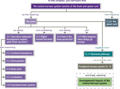
The central nervous system (CNS) consists of the brain and spinal cord. It is responsible for integrating sensory information and coordinating bodily functions. The CNS is organized into distinct regions, each with specialized roles in processing information and controlling behavior.
Brain: The main control center for processing sensory data, initiating responses, and higher mental functions.
Spinal Cord: Conducts signals to and from the brain and controls simple reflexes.
Structural Organization of the CNS
Gray Matter and White Matter
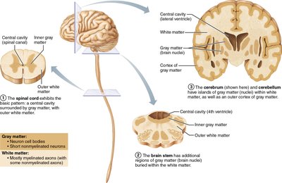
The CNS is composed of two main tissue types: gray matter and white matter. Their distribution varies between the brain and spinal cord, reflecting differences in function and connectivity.
Gray Matter: Contains neuron cell bodies, dendrites, and unmyelinated axons. It is the site of synaptic integration.
White Matter: Consists mainly of myelinated axons, which transmit signals between different CNS regions.
Spinal Cord: Central cavity surrounded by gray matter, with outer white matter.
Brain: Additional regions of gray matter (cortex and nuclei) embedded within white matter.
Ventricular System
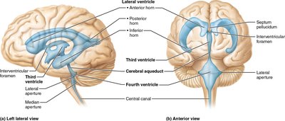
The brain contains a series of interconnected cavities called ventricles, filled with cerebrospinal fluid (CSF). These structures help protect and nourish the brain.
Lateral Ventricles: Paired structures in each cerebral hemisphere.
Third Ventricle: Located in the diencephalon, connected to lateral ventricles via interventricular foramina.
Fourth Ventricle: Located between the brainstem and cerebellum, connected to the third ventricle by the cerebral aqueduct.
Functional Areas of the Cerebral Cortex
Localization of Function
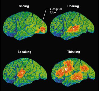
The cerebral cortex is divided into regions responsible for specific sensory, motor, and association functions. Brain imaging studies show that different activities activate distinct cortical areas.
Occipital Lobe: Visual processing (seeing).
Temporal Lobe: Auditory processing (hearing).
Frontal and Parietal Lobes: Involved in speaking and thinking.
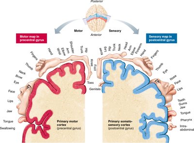
Somatotopic Maps: The Homunculus
The primary motor and somatosensory cortices are organized as maps of the body, known as homunculi. These maps reflect the amount of cortex devoted to different body regions, correlating with the complexity of movement or sensitivity.
Motor Homunculus: Located in the precentral gyrus; controls voluntary movements.
Sensory Homunculus: Located in the postcentral gyrus; processes sensory input from the body.
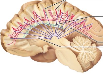
Information Flow in the Cerebral Cortex
Hierarchical Processing
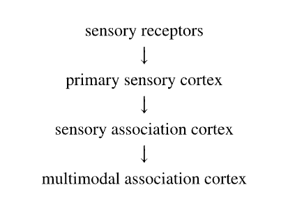
Sensory information is processed in a hierarchical manner, from initial detection to complex integration. This flow ensures that perception and response are coordinated efficiently.
Sensory Receptors → Primary Sensory Cortex: Initial detection of stimuli.
Primary Sensory Cortex → Sensory Association Cortex: Interpretation of sensory input.
Sensory Association Cortex → Multimodal Association Cortex: Integration with other sensory modalities and higher cognitive functions.
Specialized Sensory and Motor Cortices
Major Cortical Areas
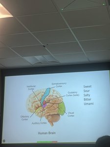
The cerebral cortex contains distinct regions for processing different types of sensory and motor information. These include:
Primary Motor Cortex: Controls voluntary movements.
Premotor Cortex: Plans and coordinates complex movements.
Broca’s Area: Involved in speech production.
Primary Somatosensory Cortex: Receives tactile information.
Visual Cortex: Processes visual input.
Auditory Cortex: Processes sound.
Vestibular Cortex: Processes balance and spatial orientation.
Olfactory Cortex: Processes smell.
Gustatory Cortex: Processes taste.
Visceral Sensory Area: Processes internal organ sensations.
Association Areas of the Cortex
Multimodal Association Areas
These regions integrate information from multiple sensory modalities and are involved in complex cognitive functions such as memory, reasoning, and personality.
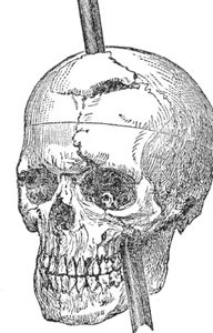
Anterior Association Area (Prefrontal Cortex): Involved in intellect, cognition, recall, and personality.
Posterior Association Area: Plays a role in recognizing patterns and faces, and integrating sensory input.
Limbic Association Area: Involved in emotional responses and memory formation.
Deeper Brain Structures
Basal Nuclei and Thalamus
The basal nuclei (ganglia) and thalamus are deep brain structures essential for motor control, sensory relay, and integration of information.
Basal Nuclei: Regulate voluntary motor activities and procedural learning.
Thalamus: Acts as a relay station for sensory information traveling to the cortex.
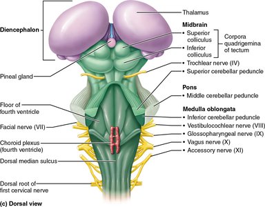
Brainstem and Cerebellum
Major Components and Functions
The brainstem connects the brain to the spinal cord and controls vital functions such as breathing and heart rate. The cerebellum coordinates movement and balance.
Midbrain, Pons, Medulla Oblongata: Control autonomic functions and relay information between the brain and spinal cord.
Cerebellum: Coordinates voluntary movements and maintains posture and balance.
Brain Waves and Sleep
Electroencephalogram (EEG) and Brain Activity
Brain waves are electrical patterns recorded from the scalp that reflect different states of brain activity. EEGs are used to study sleep, consciousness, and neurological disorders.
Alpha Waves: Awake but relaxed.
Beta Waves: Awake and alert.
Theta Waves: Common in children and during light sleep.
Delta Waves: Deep sleep.
Summary Table: Major Brain Regions and Functions
Region | Main Function(s) |
|---|---|
Cerebral Cortex | Sensory perception, voluntary movement, cognition |
Basal Nuclei | Motor control, habit learning |
Thalamus | Sensory relay to cortex |
Hypothalamus | Homeostasis, endocrine regulation |
Brainstem | Autonomic functions, cranial nerves |
Cerebellum | Coordination, balance |
Key Terms and Concepts
Gyrus: A ridge on the cerebral cortex.
Sulcus: A groove between gyri.
Association Area: Region of the cortex integrating multiple types of information.
Homunculus: A visual representation of the body mapped onto the cortex.
EEG: A test that detects electrical activity in the brain.
