 Back
The Central Nervous System: Structure and Function of the Brain and Spinal Cord
Study Guide - Smart Notes
 Tailored notes based on your materials, expanded with key definitions, examples, and context.
Brain Overview and Development
Embryological Origins and Major Brain Regions
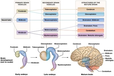
The brain and spinal cord originate from a groove on the embryonic surface, which forms the neural tube. This tube differentiates into cavities (ventricles and canals) and walls that become the mature brain's regions. The adult brain is divided into four major regions: the cerebrum, cerebellum, diencephalon, and brain stem.
Primary brain vesicles: Forebrain, midbrain, hindbrain
Secondary brain vesicles: Telencephalon, diencephalon, mesencephalon, metencephalon, myelencephalon
Structures of the mature brain: Cerebrum, diencephalon, brainstem (midbrain, pons, medulla oblongata), cerebellum
Brain Matter Organization
Gray Matter, White Matter, Nuclei, and Ganglia
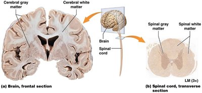
The central nervous system is organized into regions of gray matter (cell bodies, dendrites, unmyelinated axons, neuroglia) and white matter (myelinated axons in tracts). In the brain, gray matter forms the cortex, while in the spinal cord, it forms the medulla. Nuclei are clusters of gray matter within white matter, and ganglia are clusters of cell bodies outside the CNS.
Cerebrum
Structure and Functional Areas
The cerebrum is the largest brain region, responsible for higher cognitive functions. Its outermost layer, the cortex, is composed of gray matter and is known as the "thinking brain." The surface features gyri (folds), fissures (deep grooves), and sulci (shallow grooves), which help define functional areas.
Lobes of the Cerebrum
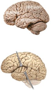
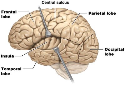
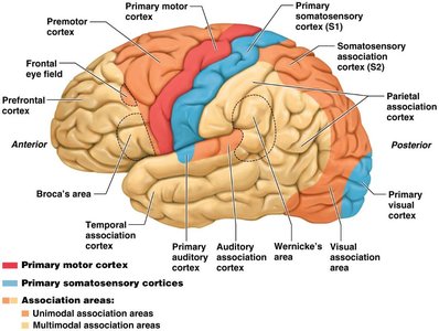
The cerebrum is divided into five lobes: frontal, parietal, temporal, occipital, and insula. The central sulcus separates the frontal and parietal lobes. The precentral gyrus is the primary motor area, and the postcentral gyrus is the primary somatosensory area.
Functional Areas of the Cerebral Cortex
The cortex contains sensory, motor, and association areas:
Sensory areas: Receive and interpret sensory input (somatosensory, visual, auditory, gustatory, olfactory)
Motor areas: Control voluntary movements (primary motor cortex, Broca's area for speech)
Association areas: Integrate information for complex functions (Wernicke's area for language comprehension)
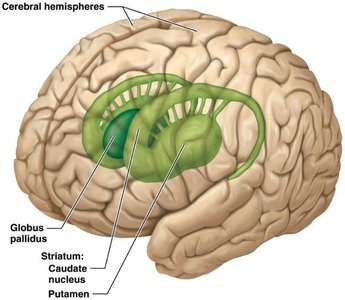
Basal Nuclei
The basal nuclei are paired masses of gray matter deep within the cerebrum. They receive input from the cortex and help initiate and terminate movements, suppress unwanted movements, regulate muscle tone, and participate in some cognitive processes.
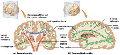
White Matter Tracts
White matter in the cerebrum consists of myelinated axons organized into tracts:
Association fibers: Connect gyri within the same hemisphere
Commissural fibers: Connect corresponding areas of the two hemispheres (e.g., corpus callosum)
Projection fibers: Connect the cerebrum with lower brain regions and the spinal cord
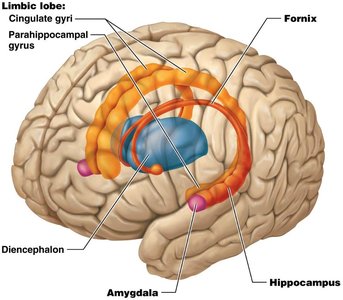
Limbic System
The limbic system is a ring of structures on the inner border of the cerebrum and floor of the diencephalon. It is involved in emotional aspects of behavior, memory, pleasure, pain, and addiction.
Diencephalon
Major Components and Functions
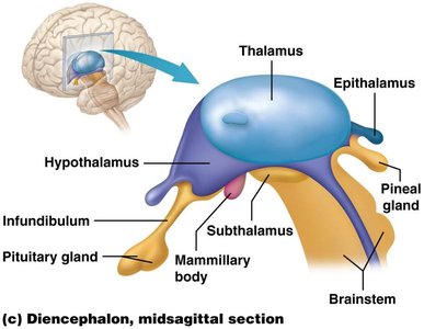
The diencephalon consists of the epithalamus, thalamus, and hypothalamus:
Epithalamus: Contains the pineal gland (secretes melatonin for diurnal cycles) and habenular nuclei (emotional responses to olfactory stimuli)
Thalamus: Relays all sensory impulses except olfaction, conscious recognition of sensation, and contributes to cognition
Hypothalamus: Primary regulator of homeostasis, controls eating, drinking, autonomic nervous system, and is associated with the pituitary gland
Cerebellum
Structure and Function
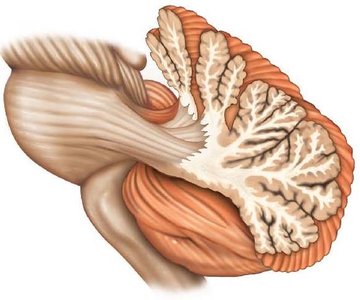
The cerebellum is the second largest brain region, attached to the brain via bundles of white matter. It regulates posture and balance, coordinates skilled voluntary movements, and compares intended versus actual movements to initiate corrections.
Brain Stem
Midbrain
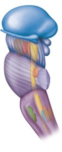
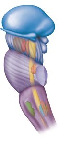
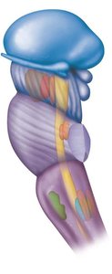
The midbrain relays motor impulses from the cerebral cortex to the pons and sensory impulses from the spinal cord to the thalamus. It coordinates movements of the eyes, head, and trunk in response to visual and auditory stimuli and contains the origin for cranial nerves III and IV.
Pons
The pons is continuous with the medulla oblongata and connects the spinal cord to the brain. It relays motor impulses from the cerebrum to the cerebellum and contains respiratory centers (pneumotaxic and apneustic areas). It is the origin for cranial nerves V-VII.
Medulla Oblongata
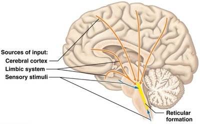
The medulla oblongata contains centers for vital reflexes (heart rate, ventilation, vasoconstriction) and non-vital reflexes (coughing, swallowing, vomiting, hiccupping, sneezing). It relays sensory and motor information between the brain and spinal cord and is the origin for cranial nerves VIII-XII.
Protection of the Brain
Cranial Bones, Meninges, and Blood-Brain Barrier
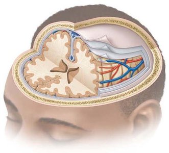
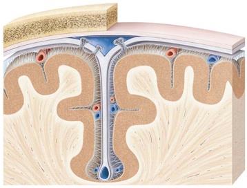
The brain is protected by cranial bones and three meninges: dura mater, arachnoid mater, and pia mater. Extensions of the dura mater (falx cerebri, falx cerebelli, tentorium cerebelli) separate parts of the brain. The blood-brain barrier prevents harmful substances and pathogens from reaching the brain, while allowing O2 and glucose to move freely.
Cerebrospinal Fluid (CSF) and Ventricles
CSF Production and Circulation
CSF is a clear, colorless liquid produced by the choroid plexus. It protects the brain and spinal cord from chemical and physical injuries and helps transport nutrients and wastes. CSF circulates through the ventricles and subarachnoid space, returning to the bloodstream via arachnoid villi.
Spinal Cord
External and Internal Anatomy
The spinal cord is protected by the vertebral column, meninges, and CSF. It extends from the medulla oblongata to approximately the L2 vertebra and has two enlargements (cervical and lumbar). There are 31 pairs of spinal nerves, named for their points of emergence.
Internal Structure and Pathways
The spinal cord's internal anatomy includes the anterior median fissure, posterior median sulcus, and central canal. Gray matter forms the medulla (anterior, posterior, lateral horns), while white matter forms the cortex (anterior, posterior, lateral columns). Sensory (ascending) tracts carry impulses to the brain, and motor (descending) tracts carry impulses from the brain.
Sensory and Motor Pathways
Sensory pathways involve first, second, and third order neurons, while motor pathways involve upper and lower motor neurons. The posterior columns/medial lemniscus system transmits proprioceptive information, while the anterolateral system (spinothalamic tracts) transmits pain, touch, and temperature. Motor commands are carried by corticospinal and corticonuclear tracts.
Central Nervous System: Homeostasis and Higher Functions
Homeostatic Regulation
The CNS regulates homeostasis through the autonomic nervous system, sleep, cardiovascular and respiratory centers, feeding, thermoregulation, and control of micturition and defecation.
Higher Mental Functions
Higher functions include localization of cognitive function, cerebral lateralization, language, learning, and memory. Brain wave activity can be measured by EEG, with different wave types corresponding to different states of consciousness and sleep stages.
