 Back
The Central Nervous System: Structure and Function
Study Guide - Smart Notes
 Tailored notes based on your materials, expanded with key definitions, examples, and context.
The Central Nervous System (CNS)
Overview
The central nervous system (CNS) consists of the brain and spinal cord. It is responsible for integrating sensory information and responding accordingly. The CNS controls vital functions such as heart rate, respiratory rate, blood pressure, and is the center for intelligence, memory, emotion, and behavior.
Rostral: Toward the nose (anterior in the brain)
Caudal: Toward the tail (posterior in the brain)
Basic Parts and Organization of the Brain
Major Regions
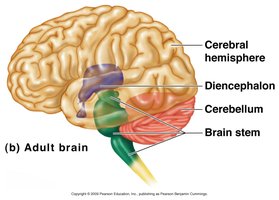
The brain is divided into four main regions: the brain stem, cerebellum, diencephalon, and cerebrum. Each region has specialized functions and structures.
Brain Stem: Includes the midbrain, pons, and medulla oblongata
Cerebellum: Coordinates movement and balance
Diencephalon: Contains the thalamus, hypothalamus, and epithalamus
Cerebrum: Largest part, responsible for higher brain functions
Brain Functions
Controls autonomic nervous system and endocrine system
Innervates the head via cranial nerves
Performs complex neural functions: intelligence, consciousness, memory, sensory-motor integration, emotion, behavior, and socialization
Ventricles of the Brain
Structure and Function
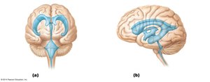
The brain contains four interconnected ventricles filled with cerebrospinal fluid (CSF). These ventricles are lined with ependymal cells and are continuous with the central canal of the spinal cord.
Lateral Ventricles: Located in the cerebral hemispheres; horseshoe-shaped
Third Ventricle: Located in the diencephalon; connected to lateral ventricles by the interventricular foramen
Cerebral Aqueduct: Connects the third and fourth ventricles
Fourth Ventricle: Located in the hindbrain; connects to the central canal of the spinal cord
Distribution of Gray and White Matter
Organization in the CNS
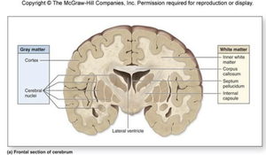
Gray matter consists of neuronal cell bodies, while white matter is composed of axons. In the brain, gray matter forms the cortex and deep nuclei, while white matter lies beneath the cortex. In the spinal cord, gray matter is central and white matter is external.
Cortex: Outer layer of gray matter in cerebrum and cerebellum
White Matter: Contains myelinated axons for communication
The Brain Stem
Structure and Function
The brain stem includes the midbrain, pons, and medulla oblongata. It acts as a passageway for all fiber tracts between the cerebrum and spinal cord and is involved in the innervation of the face and head. Ten of the twelve cranial nerves attach to the brain stem.
Produces automatic behaviors necessary for survival
Integrates auditory and visual reflexes
Medulla Oblongata
Continuous with the spinal cord
Contains pyramids (motor tracts) and inferior olivary nucleus
Four cranial nerves attach here: VIII, IX, X, XII
Contains centers for cardiac, vasomotor, and respiratory control
Pons
Bridge between midbrain and medulla oblongata
Contains nuclei for cranial nerves V, VI, VII
Connects cerebral cortex and cerebellum via middle cerebellar peduncles
Midbrain
Located between diencephalon and pons
Contains cerebral aqueduct and peduncles
Corpora quadrigemina: superior colliculi (visual reflexes), inferior colliculi (auditory reflexes)
The Cerebellum
Structure and Function
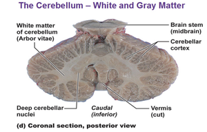
The cerebellum is located dorsal to the pons and medulla. It coordinates body movements, maintains equilibrium, and is involved in learning motor skills and cognitive functions such as language and problem-solving.
Two hemispheres, each with anterior, posterior, and flocculonodular lobes
Surface folded into folia
Composed of cortex (gray matter), arbor vitae (white matter), and deep nuclei
The Diencephalon
Major Components
The diencephalon forms the central core of the forebrain and is surrounded by the cerebral hemispheres. It consists of the thalamus, hypothalamus, and epithalamus, and borders the third ventricle.
Thalamus: Relay station for sensory information to the cerebral cortex
Hypothalamus: Main visceral control center; regulates ANS, emotions, temperature, hunger, thirst, sleep-wake cycles, and endocrine system
Epithalamus: Contains pineal gland, which secretes melatonin and regulates circadian rhythms
The Cerebrum
Structure
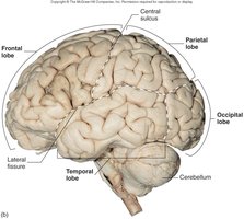
The cerebrum accounts for 83% of brain mass and is divided into lobes by deep fissures and sulci. The main lobes are frontal, parietal, occipital, temporal, and insula (deep within the lateral sulcus).
Gyri: Twisted ridges between sulci
Sulci: Grooves on the surface
Fissures: Deep grooves separating major regions
Protection of the Brain
Protective Structures
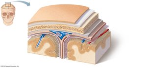
The brain is protected by the skull, meninges, cerebrospinal fluid, and the blood-brain barrier.
Meninges: Three layers—dura mater (strongest, two layers), arachnoid mater (beneath dura, contains villi for CSF absorption), pia mater (delicate, clings to brain surface)
Cerebrospinal Fluid (CSF): Cushions, nourishes, and removes waste from the brain and spinal cord; formed in choroid plexuses
Blood-Brain Barrier: Prevents most toxins from entering the brain, but allows passage of essential nutrients and some substances like alcohol and nicotine
The Spinal Cord
Structure and Function
The spinal cord provides a two-way conduction pathway between the brain and body and is a major center for reflexes. It extends from the foramen magnum to the level of vertebra L1 or L2.
Conus Medullaris: Inferior end of the spinal cord
Filum Terminale: Connective tissue attaching to the coccyx
Cervical and Lumbar Enlargements: Where nerves for limbs arise
Cauda Equina: Collection of spinal nerve roots
Gray and White Matter
Gray Matter: Central, shaped like an "H"; contains cell bodies and interneurons
White Matter: External; contains ascending, descending, and commissural fibers
Disorders of the Central Nervous System
Spinal Cord Damage
Paralysis: Loss of motor function
Parasthesia: Loss of sensation
Paraplegia: Paralysis of lower limbs (injury between T1 and L2)
Quadriplegia: Paralysis of all four limbs (injury in cervical region)
Brain Dysfunction
Cerebrovascular Accident (Stroke): Blockage or interruption of blood flow to a brain region
Alzheimer’s Disease: Progressive degenerative disease leading to dementia
Congenital Malformations
Anencephaly: Absence of cerebrum and cerebellum
Spina Bifida: Absence of vertebral lamina
Cerebral Palsy: Poor control of voluntary muscles due to cerebral cortex damage
